Drag The Labels Onto The Epidermal Layers
Holbox
Mar 19, 2025 · 5 min read
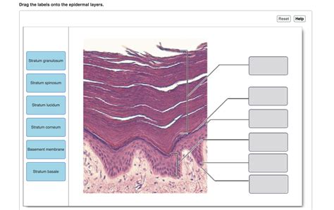
Table of Contents
Drag the Labels onto the Epidermal Layers: A Comprehensive Guide to Skin Structure and Function
Understanding the intricate layers of the epidermis is crucial for anyone studying dermatology, biology, or simply interested in skincare. This comprehensive guide dives deep into the structure and function of each epidermal layer, providing a detailed, interactive learning experience akin to a "drag the labels" exercise, but with far more depth and context.
The Epidermis: Your Body's First Line of Defense
The epidermis, the outermost layer of your skin, is a marvel of biological engineering. It's a stratified squamous epithelium, meaning it's composed of multiple layers of flattened cells. Its primary functions include:
- Protection: Acting as a barrier against harmful environmental factors like UV radiation, pathogens, and dehydration.
- Regulation: Controlling water loss and maintaining skin hydration.
- Sensation: Containing sensory receptors that transmit touch, pressure, temperature, and pain signals.
- Immune response: Participating in the immune response through the presence of Langerhans cells.
- Renewal: Constantly regenerating through a process of cell division and differentiation.
Let's explore each layer in detail, engaging in a virtual "drag the labels" exercise by mentally assigning the functions and characteristics to the appropriate stratum.
The Five Layers of the Epidermis: A Detailed Exploration
The epidermis consists of five distinct layers, each with a unique structure and function. From the deepest to the outermost, these are:
1. Stratum Basale (Germinativum): The Foundation of Renewal
(Imagine dragging the label "Cell Division" and "Melanocytes" here.)
The stratum basale is the deepest layer, resting on the basement membrane that separates the epidermis from the dermis. This is where the magic of skin regeneration begins. It's a single layer of cuboidal or columnar keratinocytes, constantly undergoing mitosis – cell division – to produce new cells. These newly formed cells are pushed upwards, slowly migrating through the other epidermal layers.
This layer also houses melanocytes, specialized cells that produce melanin, the pigment responsible for skin color and protection against UV radiation. The amount and type of melanin produced determine individual skin tone. The distribution of melanocytes is relatively uniform, with each melanocyte supplying melanin to approximately 36 keratinocytes through long, branching processes called dendrites.
Key characteristics of the stratum basale:
- High mitotic activity
- Presence of melanocytes
- Attachment to the basement membrane
- Composed of columnar or cuboidal keratinocytes
2. Stratum Spinosum: The Bridge Between Renewal and Protection
(Imagine dragging the label "Desmosomes" and "Langerhans cells" here.)
As keratinocytes migrate upwards, they enter the stratum spinosum. This layer is thicker than the stratum basale and consists of several layers of polyhedral keratinocytes. These cells appear spiny under a microscope due to the presence of numerous desmosomes, strong cell-to-cell junctions that provide structural integrity to the epidermis. The spiny appearance is an artifact of tissue preparation; in living tissue, the cells are more closely packed.
The stratum spinosum also contains Langerhans cells, specialized immune cells that act as sentinels, detecting and engulfing foreign invaders such as bacteria and viruses. They play a vital role in initiating the immune response within the skin.
Key characteristics of the stratum spinosum:
- Numerous desmosomes connecting keratinocytes
- Presence of Langerhans cells
- Increased keratinization
- Polyhedral-shaped keratinocytes
3. Stratum Granulosum: The Transition to Keratinization
(Imagine dragging the label "Keratohyalin granules" and "Lamellar granules" here.)
The stratum granulosum marks a significant transition in keratinocyte differentiation. Cells in this layer flatten and exhibit characteristic cytoplasmic granules:
- Keratohyalin granules: These granules contain proteins that are essential for the formation of keratin, the tough, fibrous protein that provides structural support to the epidermis.
- Lamellar granules: These release lipids that form a water-resistant barrier, crucial for maintaining skin hydration and preventing water loss.
The cells in this layer begin to undergo apoptosis, programmed cell death, as they continue their upward journey. This process is essential for the formation of the tough, protective outer layers of the epidermis.
Key characteristics of the stratum granulosum:
- Presence of keratohyalin granules
- Presence of lamellar granules
- Flattened keratinocytes
- Beginning of keratinization and apoptosis
4. Stratum Lucidum: A Clear Layer of Transition (Present in Thick Skin Only)
(Imagine dragging the label "Eleidin" and "Thick skin" here.)
The stratum lucidum is a thin, translucent layer found only in thick skin, such as the palms of the hands and soles of the feet. It's composed of flattened, dead keratinocytes that contain eleidin, a protein that is an intermediate stage in the conversion of keratohyalin to keratin. This layer contributes to the water-resistant properties of the thick skin.
Key characteristics of the stratum lucidum:
- Found only in thick skin
- Translucent appearance
- Contains eleidin
- Composed of flattened, dead keratinocytes
5. Stratum Corneum: The Protective Barrier
(Imagine dragging the label "Keratinized cells" and "Desquamation" here.)
The stratum corneum is the outermost layer of the epidermis, a formidable barrier composed of multiple layers of flattened, dead, keratinized cells. These cells are tightly packed together, forming a tough, protective shield against environmental insults. The keratin within these cells provides strength and resilience, while the lipids released from the lamellar granules in the stratum granulosum create the water-resistant barrier.
This layer undergoes desquamation, a continuous process of shedding dead cells, ensuring a constant renewal of the epidermal surface. This process involves the breakdown of desmosomes and the release of corneocytes.
Key characteristics of the stratum corneum:
- Composed of dead, keratinized cells
- Tough and protective
- Water-resistant barrier
- Undergoes constant desquamation
Clinical Relevance and Beyond
Understanding the structure and function of the epidermis is crucial for diagnosing and treating various skin conditions. For example, knowledge of epidermal layers helps explain the pathogenesis of conditions such as psoriasis, eczema, and skin cancers. Moreover, the development of new topical therapies and cosmetic products relies heavily on understanding how the epidermis works.
This in-depth exploration goes beyond a simple "drag the labels" exercise. It provides a comprehensive understanding of the intricate processes occurring within each epidermal layer, highlighting their interconnectivity and crucial role in maintaining skin health and overall well-being. By integrating this knowledge with practical clinical relevance, this guide provides a complete picture of the epidermis and its significance. This understanding lays a solid foundation for further learning and exploration in dermatology and related fields.
Latest Posts
Latest Posts
-
Draw The Correct Organic Product Of The Oxidation Reaction Shown
Mar 19, 2025
-
Budget Compare Actual Results To Budgeted Results
Mar 19, 2025
-
Find As A Function Of If
Mar 19, 2025
-
Where Is Velocity Highest In A River
Mar 19, 2025
-
Cups And Glasses Are Taking Too Long To Air Dry
Mar 19, 2025
Related Post
Thank you for visiting our website which covers about Drag The Labels Onto The Epidermal Layers . We hope the information provided has been useful to you. Feel free to contact us if you have any questions or need further assistance. See you next time and don't miss to bookmark.
