Correctly Label The Features Of The Larynx
Holbox
Mar 24, 2025 · 6 min read
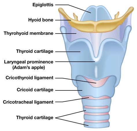
Table of Contents
- Correctly Label The Features Of The Larynx
- Table of Contents
- Correctly Labeling the Features of the Larynx: A Comprehensive Guide
- The Cartilaginous Framework: The Foundation of the Larynx
- Unpaired Cartilages:
- Paired Cartilages:
- Membranes and Ligaments: Connecting the Cartilages
- Muscles of the Larynx: Controlling Vocal Fold Movement
- Intrinsic Laryngeal Muscles:
- Extrinsic Laryngeal Muscles:
- The Glottis: The Sound-Producing Mechanism
- The Vocal Folds: Vibrating Membranes for Sound
- Neurological Control: Coordinating the Larynx
- Clinical Significance: Conditions Affecting the Larynx
- Conclusion
- Latest Posts
- Latest Posts
- Related Post
Correctly Labeling the Features of the Larynx: A Comprehensive Guide
The larynx, often referred to as the voice box, is a fascinating and complex organ. Understanding its intricate anatomy is crucial for professionals in fields like medicine, speech pathology, and singing. This comprehensive guide will delve into the detailed anatomy of the larynx, providing a clear and concise explanation of its key features and their functions. We'll ensure you can correctly label the features of the larynx with confidence.
The Cartilaginous Framework: The Foundation of the Larynx
The larynx's structure is primarily cartilaginous, providing a robust yet flexible framework. Three major unpaired cartilages and three paired cartilages form the foundation of this complex organ. Let's explore each one in detail:
Unpaired Cartilages:
-
Thyroid Cartilage: This is the largest cartilage of the larynx, shaped like a shield (hence the name "thyroid," meaning shield-shaped). It's formed by two fused laminae (plates) that meet at the midline, forming the laryngeal prominence, commonly known as the "Adam's apple." The superior border is thicker than the inferior border and the superior border is shorter than the inferior border. The thyroid cartilage is crucial for protecting the vocal folds and providing attachment points for several important muscles. Key features to label: Superior horn, inferior horn, thyroid notch, oblique line, and laminae.
-
Cricoid Cartilage: Situated inferior to the thyroid cartilage, the cricoid cartilage is a complete ring of cartilage. Its shape resembles a signet ring, with a wider posterior portion and a narrower anterior portion. Its strength and stability are essential in supporting the other laryngeal cartilages and protecting the airway. Key features to label: Posterior quadrate lamina, anterior arch.
-
Epiglottis: This leaf-shaped cartilage is located superior to the thyroid cartilage and acts as a protective flap, preventing food and liquids from entering the trachea (windpipe) during swallowing. Its flexibility allows it to move superiorly and posteriorly to cover the laryngeal inlet during swallowing. Key features to label: Petiole (stalk), body (leaf-like portion).
Paired Cartilages:
-
Arytenoid Cartilages: These pyramid-shaped cartilages are located on the superior border of the posterior cricoid cartilage. Their crucial role is in vocal fold movement and abduction (opening) and adduction (closing) of the vocal folds. They articulate with the cricoid cartilage via the cricoarytenoid joints, allowing for a wide range of movement. Key features to label: Apex, muscular process, vocal process, base.
-
Corniculate Cartilages: These small, horn-shaped cartilages sit on the apex of each arytenoid cartilage. Their function is less understood compared to the other cartilages, but they are believed to contribute to the fine adjustments of vocal fold movement. Key features to label: Their small size and location atop the arytenoids.
-
Cuneiform Cartilages: These small, rod-shaped cartilages are embedded within the aryepiglottic folds. They contribute to the support and shape of these folds, which help to protect the larynx. Key features to label: Their elongated shape and position within the aryepiglottic folds.
Membranes and Ligaments: Connecting the Cartilages
The cartilages of the larynx are interconnected by several important membranes and ligaments. These structures provide stability and facilitate movement.
-
Thyrohyoid Membrane: This membrane connects the thyroid cartilage to the hyoid bone, a U-shaped bone located superior to the larynx. It's crucial for supporting the larynx and contributing to its overall stability.
-
Cricotracheal Ligament: This ligament connects the cricoid cartilage to the first tracheal ring, providing a stable connection between the larynx and trachea.
-
Cricothyroid Ligament: This ligament connects the cricoid cartilage to the inferior border of the thyroid cartilage. This ligament is crucial as it plays an important role in adjusting vocal pitch.
Muscles of the Larynx: Controlling Vocal Fold Movement
The intricate movements of the larynx are controlled by a complex network of intrinsic and extrinsic laryngeal muscles. These muscles work together to adjust vocal fold position, tension, and length, enabling speech and other vocalizations.
Intrinsic Laryngeal Muscles:
These muscles originate and insert within the larynx itself.
-
Cricothyroid Muscle: This muscle is a key player in pitch control. By contracting, it tenses the vocal folds, resulting in higher-pitched sounds.
-
Thyroarytenoid Muscle: This muscle is a complex muscle with two main parts: the vocalis muscle and the thyromuscularis. The vocalis muscle is crucial for fine adjustments to vocal fold vibration and tone. The thyromuscularis adducts the vocal folds.
-
Posterior Cricoarytenoid Muscle: This is the only muscle responsible for abducting (opening) the vocal folds, essential for breathing.
-
Lateral Cricoarytenoid Muscle: This muscle adducts (closes) the vocal folds.
-
Transverse Arytenoid Muscle and Oblique Arytenoid Muscle: These muscles also adduct the vocal folds, further supporting the closure of the glottis.
Extrinsic Laryngeal Muscles:
These muscles originate outside the larynx and attach to it. They primarily support the larynx and influence its position. Examples include:
-
Suprahyoid Muscles: These muscles elevate the larynx, such as the digastric, stylohyoid, mylohyoid, and geniohyoid muscles.
-
Infrahyoid Muscles: These muscles depress the larynx, such as the sternohyoid, sternothyroid, omohyoid, and thyrohyoid muscles.
The Glottis: The Sound-Producing Mechanism
The glottis is the space between the vocal folds. It is the primary site of sound production. Its shape and size can change significantly, depending on the position of the vocal folds.
The Vocal Folds: Vibrating Membranes for Sound
The vocal folds (also known as vocal cords) are two mucous membrane-covered folds of tissue stretching across the larynx. Their vibration creates the sound of speech and singing. Key structures within the vocal folds include the vocal ligament and the vocalis muscle.
Key features to label: The anterior and posterior attachments of the vocal folds, the medial edge (free margin), and the lateral edge. Understanding the layered structure of the vocal folds, including the epithelium, lamina propria, and vocalis muscle, is also crucial for a complete understanding.
Neurological Control: Coordinating the Larynx
The intricate movements of the larynx are precisely coordinated by a complex network of nerves, primarily branches of the vagus nerve. The recurrent laryngeal nerve and the superior laryngeal nerve play essential roles in motor control and sensory innervation of the larynx.
Clinical Significance: Conditions Affecting the Larynx
Understanding the anatomy of the larynx is vital in diagnosing and managing a range of laryngeal conditions. Several conditions can affect the larynx, including:
- Laryngitis: Inflammation of the larynx, often causing hoarseness or voice loss.
- Laryngeal Cancer: Cancer of the larynx can affect various structures, including the vocal folds.
- Vocal Nodules: Benign growths on the vocal folds, often caused by vocal strain.
- Laryngeal Polyps: Benign growths on the vocal folds, similar to nodules.
- Paralysis of the vocal folds: Nerve damage can cause paralysis of one or both vocal folds.
Conclusion
The larynx is a marvel of biological engineering, a complex structure responsible for essential functions like breathing, swallowing, and phonation. By understanding its intricate anatomy, from the cartilaginous framework to the intricate neuromuscular control, we gain a deeper appreciation for its role in human communication and overall health. This comprehensive guide provides a robust foundation for correctly labeling the features of the larynx and understanding its crucial functions. Accurate labeling is crucial for effective communication between healthcare professionals, researchers, and educators in various fields that require understanding the complexities of the human voice box. Continue your learning with detailed anatomical atlases and further research on specific areas of interest within this complex organ.
Latest Posts
Latest Posts
-
The New Type Of Psychological Contract Has Resulted In Reduced
Mar 27, 2025
-
The Rectangular Homogeneous Gate Shown Below Is
Mar 27, 2025
-
A Clinical Trial Was Conducted To Test The Effectiveness
Mar 27, 2025
-
Murder Mystery In The Pickelson Mansion Answer Key
Mar 27, 2025
-
Is Direct Labor A Period Cost
Mar 27, 2025
Related Post
Thank you for visiting our website which covers about Correctly Label The Features Of The Larynx . We hope the information provided has been useful to you. Feel free to contact us if you have any questions or need further assistance. See you next time and don't miss to bookmark.
