All Of The Following Are Heart Valves Except
Holbox
Mar 19, 2025 · 5 min read
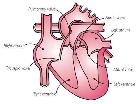
Table of Contents
All of the Following Are Heart Valves Except… Which One? A Comprehensive Guide to Cardiac Valves
The human heart, a tireless powerhouse, relies on a complex system of chambers and valves to efficiently pump blood throughout the body. Understanding these valves is crucial to grasping the mechanics of the circulatory system and recognizing potential health issues. This comprehensive guide will explore the four heart valves, their functions, and what structures are not heart valves, addressing the question: "All of the following are heart valves except…"
The Four Valves of the Heart: A Detailed Overview
The heart boasts four vital valves, each playing a unique role in ensuring unidirectional blood flow. These valves prevent backflow, crucial for maintaining efficient circulation. Let's examine each one individually:
1. Tricuspid Valve: The Right Atrium-Ventricular Gatekeeper
Located between the right atrium and the right ventricle, the tricuspid valve is so named because it possesses three cusps (leaflets) of tissue. Its primary function is to prevent the backflow of blood from the right ventricle into the right atrium during ventricular contraction (systole). This ensures that blood continues its journey towards the lungs for oxygenation. The tricuspid valve's leaflets are attached to strong tendinous cords, known as chordae tendineae, which connect to papillary muscles within the ventricle. These structures work in concert to prevent the valve leaflets from inverting under pressure.
Keywords: Tricuspid valve, right atrium, right ventricle, systole, chordae tendineae, papillary muscles, three cusps, atrioventricular valve
2. Pulmonary Valve: Guarding the Pathway to the Lungs
Positioned at the exit of the right ventricle, the pulmonary valve controls the flow of deoxygenated blood from the right ventricle into the pulmonary artery. This artery carries the blood to the lungs for gas exchange. Unlike the atrioventricular valves (tricuspid and mitral), the pulmonary valve is a semilunar valve. This means it possesses three half-moon-shaped cusps instead of the more flexible leaflets found in the atrioventricular valves. These cusps prevent the backflow of blood from the pulmonary artery into the right ventricle during diastole (relaxation).
Keywords: Pulmonary valve, right ventricle, pulmonary artery, semilunar valve, three cusps, diastole, deoxygenated blood
3. Mitral Valve: The Left Atrium-Ventricular Guardian
Situated between the left atrium and the left ventricle, the mitral valve (also known as the bicuspid valve) plays a vital role in directing oxygenated blood from the lungs into the systemic circulation. Like the tricuspid valve, it is an atrioventricular valve with two cusps. Its function is to prevent backflow from the left ventricle to the left atrium during ventricular contraction. The robust structure of the mitral valve, aided by chordae tendineae and papillary muscles, is essential for withstanding the high pressures generated by the left ventricle.
Keywords: Mitral valve, bicuspid valve, left atrium, left ventricle, atrioventricular valve, two cusps, oxygenated blood, systemic circulation
4. Aortic Valve: The Gateway to Systemic Circulation
The aortic valve, another semilunar valve, is located at the exit of the left ventricle. It controls the flow of oxygenated blood from the left ventricle into the aorta, the body's largest artery. This artery distributes oxygen-rich blood to the rest of the body. The three cusps of the aortic valve ensure that blood flows unidirectionally into the aorta and prevents regurgitation back into the left ventricle during diastole. The strong structure of this valve is crucial for handling the high pressure of systemic circulation.
Keywords: Aortic valve, left ventricle, aorta, semilunar valve, three cusps, oxygenated blood, systemic circulation, high pressure
Structures That Are NOT Heart Valves: Dispelling Common Misconceptions
Now that we've explored the four heart valves, let's address the question directly: "All of the following are heart valves except…" Many structures within the heart and surrounding vasculature might be mistaken for valves, but they serve different purposes. These include:
-
Heart Chambers (Atria and Ventricles): These are the pumping chambers of the heart, responsible for receiving and expelling blood. They do not regulate the flow of blood in the same way valves do; instead, they provide the mechanical force for blood movement.
-
Chordae Tendineae: These are fibrous cords that connect the papillary muscles to the atrioventricular valves (tricuspid and mitral). Their function is to prevent the valve leaflets from inverting during ventricular contraction, not to directly regulate blood flow. They are vital components of the valvular apparatus but are not valves themselves.
-
Papillary Muscles: These are small muscles within the ventricles. They are attached to the chordae tendineae and contract to help prevent the inversion of the atrioventricular valve leaflets during systole. While crucial for valve function, they are not valves.
-
Pulmonary Veins and Aorta: These are major blood vessels. The pulmonary veins carry oxygenated blood from the lungs to the left atrium, while the aorta distributes oxygenated blood from the left ventricle throughout the body. They transport blood but don't regulate its flow in the same way as valves.
-
Superior and Inferior Vena Cava: These large veins return deoxygenated blood from the systemic circulation to the right atrium. They are crucial for blood return, but they don't actively control blood flow direction.
-
Cardiac Conduction System: This system, comprising the sinoatrial (SA) node, atrioventricular (AV) node, Bundle of His, and Purkinje fibers, controls the heart's electrical activity and rhythm. It does not directly regulate blood flow within the heart chambers.
Clinical Significance of Heart Valve Disorders
Understanding the function and importance of each heart valve is essential for recognizing potential health problems. Valve disorders, often involving stenosis (narrowing) or regurgitation (leakage), can significantly impair heart function. These conditions can lead to various symptoms, including shortness of breath, fatigue, chest pain, and dizziness. Early diagnosis and appropriate treatment are vital for managing these conditions and improving quality of life.
Keywords: Heart valve disorders, stenosis, regurgitation, heart failure, echocardiogram, cardiac catheterization, valve replacement, valve repair
Conclusion: Understanding the Heart's Vital Valves
This exploration of the heart's valves highlights their critical role in maintaining efficient and unidirectional blood flow. We’ve clarified the distinct functions of the tricuspid, pulmonary, mitral, and aortic valves, contrasting them with other cardiac structures. Recognizing the differences is key to comprehending the intricate workings of the cardiovascular system and appreciating the implications of valve dysfunction. Remembering the functions of these four vital valves and the structures that are not valves is crucial for both healthcare professionals and those seeking a deeper understanding of the human body. Further research into specific valve disorders and their treatments can provide even greater insight into this complex and fascinating system.
Latest Posts
Latest Posts
-
If The Supply Of Green Tea Rises
Mar 19, 2025
-
Umatilla Bank And Trust Is Considering Giving
Mar 19, 2025
-
An Ordinary Annuity Is Best Defined As
Mar 19, 2025
-
A Flexible Budget Performance Report Compares
Mar 19, 2025
-
Standard Heat Of Formation For H2o
Mar 19, 2025
Related Post
Thank you for visiting our website which covers about All Of The Following Are Heart Valves Except . We hope the information provided has been useful to you. Feel free to contact us if you have any questions or need further assistance. See you next time and don't miss to bookmark.
