Where Is The Tissue Pictured Found
Holbox
Mar 17, 2025 · 6 min read
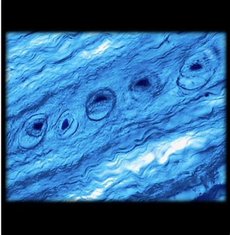
Table of Contents
Where is the Tissue Pictured Found? A Comprehensive Guide to Tissue Localization
Finding the precise location of a pictured tissue requires a multi-faceted approach, combining visual analysis with a deep understanding of histology and anatomy. This article delves into the methodologies used to identify tissue origin from images, highlighting the crucial role of microscopic features, staining techniques, and the overall context provided within the image. We'll explore various scenarios, discussing limitations and offering strategies to improve the accuracy of your tissue localization.
Understanding the Challenges of Tissue Identification
Before diving into the techniques, it's vital to acknowledge the inherent difficulties in identifying tissue solely from a picture. Several factors contribute to this complexity:
-
Image Resolution and Quality: Low-resolution images or those with poor contrast significantly hinder accurate identification. Fine details crucial for tissue differentiation might be lost.
-
Staining Techniques: Different staining methods (e.g., H&E, PAS, immunohistochemistry) highlight different cellular components, leading to vastly different visual appearances of the same tissue. Identifying the staining technique used is paramount.
-
Magnification: The magnification level greatly influences the observable details. A low-magnification view might show overall tissue architecture, while high magnification reveals cellular features.
-
Artifacts: Processing and imaging artifacts can introduce distortions, making the interpretation challenging. These artifacts could mimic pathological changes or mask true tissue characteristics.
-
Partial Views: Images often show only a small section of a larger tissue sample, hindering a complete understanding of its location and context.
A Systematic Approach to Tissue Localization
To effectively determine the tissue's origin, follow a methodical approach:
1. Analyze Microscopic Features:
-
Cell Morphology: Carefully examine the shape, size, and arrangement of cells. Epithelial cells, for example, tend to be tightly packed, while connective tissue cells are more dispersed. The presence of specialized cells (e.g., neurons, muscle fibers) provides strong clues.
-
Extracellular Matrix (ECM): The composition and arrangement of the ECM are highly tissue-specific. Observe the presence of collagen fibers, elastic fibers, ground substance, and other components. The staining method will significantly influence the visualization of the ECM.
-
Tissue Organization: Notice the overall tissue arrangement – is it stratified, columnar, or cuboidal epithelium? Is it organized into layers or bundles? This organizational pattern is highly diagnostic.
-
Special Structures: Identify any unique structures present, such as glands, blood vessels, hair follicles, or nerve bundles. The presence of specific structures significantly narrows down the possibilities.
2. Identify the Staining Technique:
-
Hematoxylin and Eosin (H&E): The most common staining technique, H&E stains nuclei purple/blue and cytoplasm pink/red. This allows for basic cell identification.
-
Periodic Acid-Schiff (PAS): PAS stains carbohydrates, particularly glycogen and glycoproteins, magenta. This is useful for identifying structures rich in carbohydrates, like mucus-secreting cells.
-
Trichrome stains: These stains differentiate collagen from other tissue components, providing information about connective tissue composition.
-
Immunohistochemistry (IHC): IHC employs antibodies to target specific proteins. The staining pattern reveals the presence or absence of specific proteins, providing highly specific information about the tissue type.
3. Consider the Contextual Information:
-
Image Caption or Metadata: Always check for any accompanying information, such as the source of the image, the tissue preparation method, and any clinical details.
-
Surrounding Tissues: If the image shows adjacent tissues, this significantly aids in localization. The relationship between different tissues often provides crucial information.
-
Gross Anatomy: If a macroscopic image of the organ is available, this provides an excellent anatomical context, narrowing down the possible locations within the organ itself.
Specific Examples and Their Localization Strategies
Let's consider some specific tissue examples and discuss how you'd approach their localization:
A. Epithelial Tissue:
To pinpoint the location of an epithelial tissue pictured, consider:
-
Type of Epithelium: Is it stratified squamous, simple columnar, pseudostratified columnar, or transitional epithelium? Each type is found in specific locations.
-
Location of Basal Lamina: Identifying the basal lamina, the supportive layer beneath epithelium, helps determine the interface with underlying connective tissue.
-
Associated Structures: Glands, cilia, microvilli, and keratinization (in stratified squamous epithelium) provide additional clues.
Stratified squamous epithelium, for example, is found in the epidermis of the skin, the lining of the esophagus, and the vagina. Its location is determined by the presence of keratinization and the absence of other specialized structures.
B. Connective Tissue:
Identifying the precise location of connective tissue requires focusing on:
-
Fiber Type and Arrangement: Collagen fibers are abundant in many connective tissues, while elastic fibers are more prevalent in tissues requiring flexibility. Their arrangement (e.g., parallel, interwoven) further informs localization.
-
Cell Types: Fibroblasts, adipocytes, chondrocytes, and osteocytes are characteristic of different connective tissues.
-
Matrix Composition: The ground substance varies between different connective tissues, impacting the tissue's overall properties and location.
For example, dense regular connective tissue, with its parallel collagen fibers, is found in tendons and ligaments. Adipose tissue, characterized by its abundance of adipocytes, is found in subcutaneous layers and around organs.
C. Muscle Tissue:
Determining the location of muscle tissue requires analyzing:
-
Muscle Fiber Type: Skeletal muscle fibers are striated and multinucleated, while smooth muscle fibers are non-striated and uninucleated. Cardiac muscle fibers are striated and branched, with intercalated discs.
-
Arrangement of Fibers: The arrangement of muscle fibers provides crucial information. For example, skeletal muscle fibers are often arranged in parallel bundles.
-
Associated Structures: The presence of tendons, nerves, and blood vessels helps determine the muscle's location and function.
Skeletal muscle is attached to bones, while smooth muscle is found in the walls of internal organs. Cardiac muscle is located exclusively in the heart.
D. Nervous Tissue:
Identifying the location of nervous tissue depends on:
-
Neuron Morphology: The shape and size of neurons vary widely depending on their location and function.
-
Glial Cells: The presence and type of glial cells (e.g., astrocytes, oligodendrocytes) further assist localization.
-
Myelin Sheaths: The presence of myelin sheaths indicates myelinated axons, commonly found in the white matter of the brain and spinal cord.
The brain and spinal cord are the primary locations of nervous tissue, but nervous tissue also extends throughout the peripheral nervous system.
Limitations and Considerations
It's crucial to acknowledge that even with meticulous analysis, accurately identifying tissue location from a single image can be challenging. Multiple images from different angles and magnifications are highly recommended. Consulting with a pathologist or histologist significantly improves the accuracy of the identification. Finally, remember that context is king. The more information you have, the greater your chances of accurately determining the tissue's origin.
By combining visual analysis, a thorough understanding of histology and anatomy, and a systematic approach, you can significantly increase your ability to determine the location of a pictured tissue. Always be mindful of the limitations, and consult with experts when necessary to ensure accuracy. Remember that this process requires careful observation, and practice will sharpen your skills.
Latest Posts
Latest Posts
-
It Is Unusual For A Company To Sell
Mar 17, 2025
-
The Impact Of Technology On Internal Controls Includes
Mar 17, 2025
-
For The Substituted Cyclohexane Compound Shown
Mar 17, 2025
-
Select The Correct Statement About The Regulation Of Gastric Secretion
Mar 17, 2025
-
Which Strategy Teaches Healthier Ways To Use Substances
Mar 17, 2025
Related Post
Thank you for visiting our website which covers about Where Is The Tissue Pictured Found . We hope the information provided has been useful to you. Feel free to contact us if you have any questions or need further assistance. See you next time and don't miss to bookmark.
