Use The Key On The Right To Identify The Arteries
Holbox
Mar 22, 2025 · 7 min read
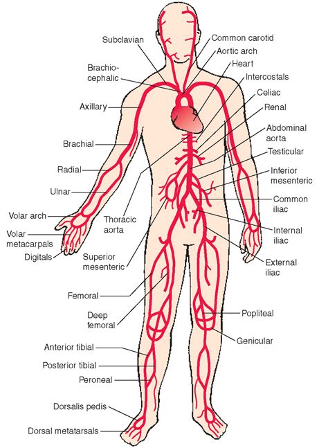
Table of Contents
- Use The Key On The Right To Identify The Arteries
- Table of Contents
- Use the Key on the Right to Identify the Arteries: A Comprehensive Guide to Arterial Anatomy
- Understanding Arterial Anatomy: A Foundation for Identification
- Key Features for Arterial Identification:
- Identifying Arteries of the Head and Neck: A Regional Approach
- 1. Common Carotid Artery:
- 2. Internal Carotid Artery:
- 3. External Carotid Artery:
- Arteries of the Thorax: Fueling Vital Organs
- 1. Aorta:
- 2. Coronary Arteries:
- 3. Pulmonary Arteries:
- Abdominal Arteries: Supplying the Digestive System and More
- 1. Abdominal Aorta:
- 2. Celiac Trunk:
- 3. Superior Mesenteric Artery:
- 4. Renal Arteries:
- 5. Inferior Mesenteric Artery:
- Arteries of the Upper and Lower Limbs: Reaching the Extremities
- Upper Limb Arteries:
- Lower Limb Arteries:
- Using a Key for Arterial Identification: Practical Application
- Importance of Accurate Arterial Identification: Clinical Significance
- Conclusion: Mastering Arterial Anatomy for Enhanced Understanding
- Latest Posts
- Latest Posts
- Related Post
Use the Key on the Right to Identify the Arteries: A Comprehensive Guide to Arterial Anatomy
Identifying arteries correctly is crucial for medical professionals, anatomy students, and anyone interested in understanding the human circulatory system. This comprehensive guide will walk you through the process of identifying major arteries using a key, focusing on anatomical location, branching patterns, and associated structures. We'll cover key arteries of the head, neck, thorax, abdomen, and limbs, providing detailed descriptions and visual aids (although images themselves cannot be included within this text-based format). Remember, always refer to detailed anatomical atlases and diagrams for accurate visualization.
Understanding Arterial Anatomy: A Foundation for Identification
Before diving into artery identification, it's vital to grasp fundamental concepts of arterial anatomy. Arteries are blood vessels that carry oxygenated blood away from the heart. They are characterized by their thick, elastic walls, which enable them to withstand the high pressure of blood ejected from the heart. The arterial system is a complex network, branching repeatedly to deliver oxygen and nutrients to all parts of the body. Understanding this branching pattern is key to accurate identification.
Key Features for Arterial Identification:
- Location: The anatomical position of an artery is often the first clue to its identity. Knowing the general region (e.g., neck, arm, leg) significantly narrows down the possibilities.
- Branching Pattern: Arteries frequently branch into smaller vessels. The pattern of branching—whether it's symmetrical, asymmetrical, or forms a specific network—is a crucial identifier.
- Associated Structures: Arteries often run alongside nerves, veins, or other anatomical structures. These relationships provide further clues to identification.
- Pulse: Palpable pulses can help locate major arteries near the surface of the skin.
- Size and Diameter: While not always easily discernible visually, the relative size of an artery can be helpful in identifying larger versus smaller vessels.
Identifying Arteries of the Head and Neck: A Regional Approach
The arteries supplying the head and neck are branches of the common carotid arteries, which bifurcate into the internal and external carotid arteries.
1. Common Carotid Artery:
- Location: Runs along the side of the neck, deep to the sternocleidomastoid muscle.
- Branches: Bifurcates into the internal and external carotid arteries.
- Identification: Palpable pulse in the neck.
2. Internal Carotid Artery:
- Location: Enters the skull through the carotid canal.
- Branches: Supplies the brain and orbits. Includes the ophthalmic artery, anterior cerebral artery, and middle cerebral artery (among others).
- Identification: Not directly palpable; its branches can be identified through angiography.
3. External Carotid Artery:
- Location: Ascends through the neck, branching extensively to supply the face, scalp, and neck.
- Branches: Includes the superior thyroid artery, lingual artery, facial artery, occipital artery, posterior auricular artery, superficial temporal artery, and maxillary artery.
- Identification: Several branches are palpable (e.g., facial artery at the angle of the mandible).
Arteries of the Thorax: Fueling Vital Organs
The arteries of the thorax primarily originate from the aorta, the body's largest artery.
1. Aorta:
- Location: Arises from the left ventricle of the heart. It has ascending, aortic arch, and descending portions.
- Branches: Numerous branches supply the heart, lungs, and other thoracic structures. Key branches include the coronary arteries, brachiocephalic trunk, left common carotid artery, left subclavian artery, and intercostal arteries.
- Identification: Not directly palpable; its branches are identifiable through imaging techniques.
2. Coronary Arteries:
- Location: Supply the heart muscle itself.
- Branches: Right and left coronary arteries, each branching extensively.
- Identification: Visualized through coronary angiography.
3. Pulmonary Arteries:
- Location: Carry deoxygenated blood from the right ventricle to the lungs.
- Branches: Right and left pulmonary arteries, each branching into smaller arteries within the lungs.
- Identification: Visible during thoracic surgery or imaging procedures.
Abdominal Arteries: Supplying the Digestive System and More
The abdominal aorta is a continuation of the thoracic aorta, giving rise to several major arteries that supply the abdominal viscera and lower limbs.
1. Abdominal Aorta:
- Location: Descends through the abdomen, posterior to the abdominal viscera.
- Branches: Celiac trunk, superior mesenteric artery, renal arteries, inferior mesenteric artery, and common iliac arteries (among many others).
- Identification: Visible during abdominal surgery or imaging procedures.
2. Celiac Trunk:
- Location: Branches off the abdominal aorta, supplying the stomach, liver, spleen, and pancreas.
- Branches: Left gastric artery, splenic artery, and common hepatic artery.
- Identification: Visualized during abdominal surgery or angiography.
3. Superior Mesenteric Artery:
- Location: Supplies the small intestine and part of the large intestine.
- Branches: Numerous small arteries supplying the intestinal loops.
- Identification: Visible during abdominal surgery or angiography.
4. Renal Arteries:
- Location: Supply the kidneys.
- Branches: One for each kidney.
- Identification: Visible during abdominal surgery or angiography.
5. Inferior Mesenteric Artery:
- Location: Supplies the distal large intestine.
- Branches: Left colic artery, sigmoid arteries, and superior rectal artery.
- Identification: Visible during abdominal surgery or angiography.
Arteries of the Upper and Lower Limbs: Reaching the Extremities
The arteries supplying the upper and lower limbs originate from the subclavian and common iliac arteries, respectively.
Upper Limb Arteries:
-
Subclavian Artery: Continues as the axillary artery, then brachial artery, branching into the radial and ulnar arteries. These further subdivide to form the palmar arches and digital arteries. Identification involves palpation of pulses (brachial, radial, ulnar) and using anatomical landmarks.
-
Axillary Artery: Located in the armpit, it gives off branches to the shoulder and chest.
-
Brachial Artery: Runs along the medial aspect of the arm, providing blood to the muscles and tissues of the arm. It's easily palpated at the elbow.
-
Radial and Ulnar Arteries: These arteries run along the forearm. The radial pulse is commonly used for pulse checks.
Lower Limb Arteries:
-
Common Iliac Arteries: These bifurcate into the external and internal iliac arteries. The external iliac artery becomes the femoral artery.
-
Femoral Artery: Located in the thigh, this artery is a major blood supply to the leg and provides readily palpable femoral pulse.
-
Popliteal Artery: Located behind the knee, giving off branches to the knee joint.
-
Anterior and Posterior Tibial Arteries: These arteries supply the muscles and tissues of the lower leg and foot, with palpable pulses at the dorsalis pedis and posterior tibial arteries.
Using a Key for Arterial Identification: Practical Application
A key typically presents a series of dichotomous choices based on anatomical location, branching patterns, and associated structures. For example:
Key:
1a. Artery located in the neck: Go to 2 1b. Artery located in the arm: Go to 7 ...and so on...
2a. Artery bifurcates into internal and external carotid arteries: Common Carotid Artery 2b. Artery supplies the brain: Internal Carotid Artery ...and so on...
By systematically working through the key, you can progressively narrow down the possibilities until you arrive at the correct identification. Remember that using a key requires a strong understanding of arterial anatomy and a familiarity with the terminology used in the key itself.
Importance of Accurate Arterial Identification: Clinical Significance
Accurate identification of arteries is of paramount importance in various medical contexts:
- Surgery: Precise knowledge of arterial anatomy is crucial during surgical procedures to avoid accidental injury to major blood vessels.
- Angiography: Angiography, a medical imaging technique, relies on the accurate identification of arteries to visualize blood flow and detect blockages.
- Trauma Care: In emergency situations involving trauma, rapid and accurate identification of arteries is essential for controlling bleeding.
- Medical Education: Thorough understanding of arterial anatomy is a cornerstone of medical training.
Conclusion: Mastering Arterial Anatomy for Enhanced Understanding
Mastering the art of identifying arteries requires diligent study, practice, and a solid understanding of anatomical principles. By combining knowledge of anatomical location, branching patterns, associated structures, and the use of identification keys, you can confidently navigate the intricate network of the arterial system. Remember to always cross-reference your findings with reliable anatomical resources to ensure accuracy. This guide provides a strong foundation, but continuous learning and practical experience are key to becoming proficient in arterial identification. Always prioritize safety and consult with qualified professionals when dealing with human anatomy.
Latest Posts
Latest Posts
-
When Calcium Ions Enter The Synaptic Terminal
Mar 24, 2025
-
Which Of The Following Is Equivalent To
Mar 24, 2025
-
Essentials Of Cultural Anthropology A Toolkit For A Global Age
Mar 24, 2025
-
Correctly Label The Features Of The Larynx
Mar 24, 2025
-
A Large Sunflower Population Is Established In A Field
Mar 24, 2025
Related Post
Thank you for visiting our website which covers about Use The Key On The Right To Identify The Arteries . We hope the information provided has been useful to you. Feel free to contact us if you have any questions or need further assistance. See you next time and don't miss to bookmark.
