The Knee Jerk Reflex Is An Example Of A
Holbox
Mar 19, 2025 · 7 min read
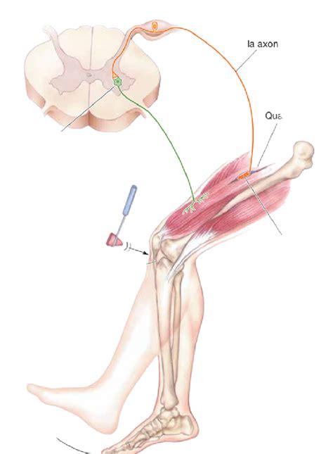
Table of Contents
The Knee-Jerk Reflex: A Prime Example of a Monosynaptic Reflex Arc
The knee-jerk reflex, also known as the patellar reflex, is a classic example of a monosynaptic reflex arc. This seemingly simple reaction to a tap below the kneecap reveals a complex interplay of neural pathways and muscle actions, providing a fascinating window into the workings of the human nervous system. Understanding the knee-jerk reflex is crucial for comprehending fundamental neurological principles, diagnosing neurological disorders, and appreciating the intricate mechanisms of human movement.
Understanding Reflex Arcs: The Body's Automatic Responses
Before diving into the specifics of the knee-jerk reflex, let's establish a basic understanding of reflex arcs. A reflex arc is a neural pathway that mediates a reflex action. Reflexes are involuntary, rapid, and predictable responses to stimuli. They bypass the brain's higher processing centers, ensuring swift reactions to potentially harmful situations. This rapid response is crucial for survival; imagine the delay if your hand had to wait for your brain to fully process the sensation of touching a hot stove before initiating a withdrawal!
Reflex arcs typically consist of the following components:
-
Receptor: This specialized structure detects the stimulus. In the case of the knee-jerk reflex, the receptor is a muscle spindle within the quadriceps muscle. Muscle spindles are sensitive to changes in muscle length and stretch.
-
Sensory Neuron (Afferent Neuron): This neuron transmits the sensory information from the receptor to the central nervous system (CNS), which in this case is the spinal cord. The sensory neuron's axon enters the spinal cord through the dorsal root.
-
Integration Center: This is the area within the CNS where the sensory information is processed. In monosynaptic reflexes, like the knee-jerk reflex, the integration center is remarkably simple – a single synapse between the sensory neuron and the motor neuron.
-
Motor Neuron (Efferent Neuron): This neuron carries the motor command from the CNS to the effector. Its axon exits the spinal cord via the ventral root.
-
Effector: This is the muscle or gland that carries out the response. In the knee-jerk reflex, the effector is the quadriceps muscle, responsible for extending the leg.
The Monosynaptic Nature of the Knee-Jerk Reflex
The knee-jerk reflex is classified as monosynaptic because it involves only one synapse between the sensory and motor neuron. This direct connection allows for an exceptionally fast response time, typically around 30-50 milliseconds. The simplicity of this pathway is key to its speed and efficiency. Contrast this with polysynaptic reflexes, which involve multiple synapses and interneurons, resulting in a more complex and slower response.
The Detailed Mechanism: A Step-by-Step Breakdown
-
Stimulus: A sharp tap on the patellar tendon below the kneecap stretches the quadriceps muscle.
-
Receptor Activation: This stretch activates the muscle spindles within the quadriceps, triggering the generation of nerve impulses.
-
Sensory Neuron Activation: These impulses travel along the sensory neuron's axon to the spinal cord.
-
Synapse at the Spinal Cord: The sensory neuron directly synapses with the motor neuron in the spinal cord's gray matter. This is the single synapse that defines the reflex as monosynaptic. No interneurons are involved in this simple pathway.
-
Motor Neuron Activation: The neurotransmitter, acetylcholine, is released at the synapse, stimulating the motor neuron.
-
Effector Response: The motor neuron's impulse travels along its axon to the quadriceps muscle.
-
Muscle Contraction: Acetylcholine is released at the neuromuscular junction, causing the quadriceps muscle to contract, extending the lower leg.
Inhibitory Component: The Role of Reciprocal Innervation
While the knee-jerk reflex primarily involves the contraction of the quadriceps, it also incorporates an inhibitory component involving the reciprocal innervation of the hamstring muscles. Simultaneously with the excitation of the quadriceps motor neurons, inhibitory interneurons are activated. These interneurons synapse with motor neurons that innervate the hamstring muscles, the antagonists of the quadriceps. This inhibition of the hamstrings prevents them from opposing the extension of the leg, ensuring a smooth and efficient response. This coordinated action exemplifies the finely tuned control of muscle activity within the nervous system.
Clinical Significance: Assessing Neurological Function
The knee-jerk reflex test is a fundamental part of neurological examinations. Its ease of elicitation and straightforward assessment makes it a valuable tool for evaluating the integrity of the spinal cord, peripheral nerves, and neuromuscular junctions. An absent or diminished reflex (hyporeflexia) or an exaggerated reflex (hyperreflexia) can indicate underlying neurological issues, such as:
-
Lower motor neuron lesions: Damage to the motor neuron or its axon, such as in peripheral neuropathy or poliomyelitis, can lead to hyporeflexia or areflexia (complete absence of the reflex).
-
Upper motor neuron lesions: Damage to the upper motor neuron pathways, such as in stroke or multiple sclerosis, can result in hyperreflexia, often accompanied by other neurological signs like spasticity and clonus.
-
Metabolic disorders: Conditions affecting electrolyte balance, such as hypokalemia or hypothyroidism, can influence reflex activity.
-
Muscular diseases: Myopathies, such as muscular dystrophy, can impair the muscle's response to neuronal stimulation, resulting in hyporeflexia.
The careful evaluation of the knee-jerk reflex, along with other neurological tests, is essential in diagnosing and managing a wide range of neurological conditions. The intensity of the reflex, its symmetry between the two legs, and the presence of any associated abnormal movements are all crucial elements in clinical interpretation.
Beyond the Basics: Factors Influencing the Knee-Jerk Reflex
Several factors can influence the intensity and speed of the knee-jerk reflex:
-
Jendrassik maneuver: This technique involves clenching the fists and pulling the hands apart, which enhances the reflex response by increasing the overall level of neural activity in the spinal cord. This illustrates how descending pathways from the brain can modulate reflex activity.
-
Muscle fatigue: Fatigue in the quadriceps muscle can weaken the reflex response.
-
Attention and mental state: Although reflexes are generally considered involuntary, conscious efforts to suppress or enhance the reflex have been shown to have some influence on its intensity.
-
Age: The intensity of the reflex tends to decrease with age.
The Knee-Jerk Reflex: A Window into Neurological Function
The seemingly simple knee-jerk reflex provides a powerful illustration of the complexity and precision of the nervous system. Its monosynaptic nature allows for an extremely rapid response, crucial for maintaining posture and balance. Furthermore, its clinical significance makes it an invaluable tool in neurological diagnosis. By understanding the intricacies of this fundamental reflex, we gain deeper insights into the intricate mechanisms that govern human movement and the remarkable adaptability of our nervous system. The next time you have your reflexes tested, remember the detailed process occurring at the level of your spinal cord, a testament to the sophistication of the human body's built-in defense mechanisms. The simple tap on your knee reveals a world of complex neurological processes working in perfect harmony.
Further Exploration: Related Reflexes and Neurological Concepts
While the knee-jerk reflex is a prime example of a monosynaptic reflex, the human body utilizes a wide array of other reflexes, both monosynaptic and polysynaptic, to maintain homeostasis and respond to environmental stimuli. Exploring these related concepts expands our understanding of the nervous system's remarkable capabilities:
-
Other Stretch Reflexes: The knee-jerk reflex is just one example of a stretch reflex, a crucial mechanism for maintaining muscle tone and posture. Other stretch reflexes include the biceps reflex, triceps reflex, and Achilles reflex.
-
Withdrawal Reflex: This polysynaptic reflex protects the body from harmful stimuli, such as heat or pain. It involves the coordinated withdrawal of a limb from the source of the stimulus.
-
Flexor Reflex: This reflex, often occurring in conjunction with the withdrawal reflex, involves the contraction of flexor muscles to withdraw a limb from a painful stimulus.
-
Crossed Extensor Reflex: This reflex, usually paired with a flexor reflex, stabilizes the body during a withdrawal reflex by extending the opposite limb. For example, if you step on a sharp object, the flexor reflex will pull your injured leg away, while the crossed extensor reflex will extend the opposite leg to maintain balance.
By delving deeper into these related reflex arcs and neurological principles, a richer and more comprehensive picture of the body's amazing ability to respond automatically to its environment emerges. This reinforces the significance of the knee-jerk reflex as a foundational element in understanding the complexities of the human nervous system.
Latest Posts
Latest Posts
-
A Coffee Producer Has Two Social Media Objectives
Mar 19, 2025
-
What Is The Medial Border Of The Highlighted Region Called
Mar 19, 2025
-
Select The Account Classification That Matches With The Description
Mar 19, 2025
-
An Inbound Sales Rep For A Digital
Mar 19, 2025
-
A Firms Supply Curve Is Upsloping Because
Mar 19, 2025
Related Post
Thank you for visiting our website which covers about The Knee Jerk Reflex Is An Example Of A . We hope the information provided has been useful to you. Feel free to contact us if you have any questions or need further assistance. See you next time and don't miss to bookmark.
