Spotlight Figure 10.10 Neuromuscular Junction Nmj
Holbox
Mar 19, 2025 · 6 min read
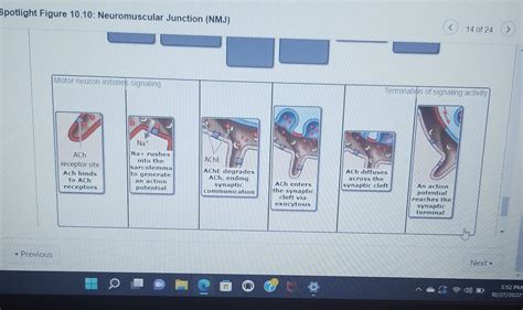
Table of Contents
Spotlight on Figure 10.10: Neuromuscular Junction (NMJ) – A Deep Dive into the Exquisite Dance of Nerve and Muscle
Figure 10.10, often found in neuroscience textbooks, depicts the neuromuscular junction (NMJ), a fascinating and intricate structure crucial for voluntary movement. This detailed illustration unveils the exquisite choreography between a motor neuron and a muscle fiber, a communication pathway vital for our everyday actions, from the simplest twitch to the most complex athletic feats. This article will delve into the intricacies of Figure 10.10, exploring the key components, the mechanisms of neurotransmission, and the significance of the NMJ in health and disease.
Understanding the Anatomy of the Neuromuscular Junction (NMJ)
The NMJ, also known as the myoneural junction, is a specialized synapse where a motor neuron transmits a signal to a skeletal muscle fiber, initiating muscle contraction. Figure 10.10 typically showcases several key anatomical features:
1. The Presynaptic Terminal (Motor Neuron Axon Terminal):
This is the endpoint of the motor neuron axon. It's characterized by a swollen appearance, packed with numerous synaptic vesicles. These vesicles are tiny sacs brimming with acetylcholine (ACh), a crucial neurotransmitter for muscle contraction. The presynaptic terminal's integrity is paramount; any damage can lead to impaired neurotransmission and muscle weakness. Figure 10.10 likely highlights the presence of voltage-gated calcium channels (Ca²⁺ channels) within the presynaptic membrane. These channels are essential for triggering the release of ACh. When an action potential reaches the presynaptic terminal, it depolarizes the membrane, opening these channels, allowing calcium ions to flood into the terminal.
2. The Synaptic Cleft:
This is the narrow gap separating the presynaptic terminal from the postsynaptic membrane of the muscle fiber. It's a meticulously controlled space, approximately 20-30 nanometers wide. The synaptic cleft ensures the neurotransmitter's targeted delivery to the muscle fiber, preventing widespread activation of other tissues. The composition of the synaptic cleft influences the speed and efficiency of neurotransmission. Figure 10.10 might indicate the presence of various enzymes and molecules in this space, such as acetylcholinesterase (AChE), responsible for degrading ACh and regulating the duration of the muscle contraction.
3. The Postsynaptic Membrane (Motor End Plate):
This specialized region of the muscle fiber membrane lies opposite the presynaptic terminal. It's characterized by a high density of acetylcholine receptors (AChRs), transmembrane proteins that bind ACh. These receptors are ligand-gated ion channels, meaning they open when ACh binds, allowing the influx of sodium ions (Na⁺) into the muscle fiber. This influx of sodium ions causes a depolarization, generating an end-plate potential (EPP). The high concentration of AChRs ensures efficient signal transmission and a strong muscle response. Figure 10.10 likely emphasizes the folds and creases in the postsynaptic membrane, which significantly increase the surface area available for ACh binding and amplify the signal. These junctional folds are a defining characteristic of the NMJ.
4. Supporting Cells:
While not always prominently displayed, Figure 10.10 may subtly suggest the presence of supporting cells, such as Schwann cells. These cells wrap around the axon terminal and play a vital role in maintaining the health and proper functioning of the NMJ. They provide structural support, contribute to the formation of the synaptic cleft, and modulate neurotransmitter release.
The Mechanism of Neuromuscular Transmission: A Step-by-Step Guide
The transmission of a nerve impulse from the motor neuron to the muscle fiber is a precisely orchestrated sequence of events. Let's examine the process using Figure 10.10 as our guide:
-
Nerve Impulse Arrival: An action potential (nerve impulse) travels down the motor neuron axon and reaches the presynaptic terminal.
-
Calcium Influx: The depolarization of the presynaptic terminal opens voltage-gated calcium channels, allowing calcium ions (Ca²⁺) to enter the terminal.
-
Vesicle Fusion and Neurotransmitter Release: The influx of calcium ions triggers the fusion of synaptic vesicles with the presynaptic membrane, releasing ACh into the synaptic cleft. This process is known as exocytosis.
-
Acetylcholine Binding: ACh diffuses across the synaptic cleft and binds to AChRs on the postsynaptic membrane of the muscle fiber.
-
Ion Channel Opening and Depolarization: ACh binding opens the ligand-gated ion channels, allowing sodium ions (Na⁺) to rush into the muscle fiber. This influx of positive charges depolarizes the postsynaptic membrane, generating the end-plate potential (EPP).
-
Muscle Fiber Excitation: The EPP triggers the opening of voltage-gated sodium channels along the muscle fiber membrane, initiating an action potential that spreads throughout the muscle fiber.
-
Muscle Contraction: The action potential in the muscle fiber leads to the release of calcium ions from the sarcoplasmic reticulum (SR), initiating the sliding filament mechanism of muscle contraction.
-
Acetylcholine Degradation: Acetylcholinesterase (AChE) in the synaptic cleft quickly breaks down ACh, terminating the signal and preventing prolonged muscle contraction. This rapid degradation is critical for precise control of muscle movement.
The Significance of the Neuromuscular Junction in Health and Disease
The NMJ's proper functioning is essential for normal muscle activity. Disruptions at the NMJ can lead to a variety of neuromuscular disorders, many of which are debilitating. Figure 10.10 provides a visual foundation for understanding these disorders:
1. Myasthenia Gravis:
This autoimmune disease targets AChRs, leading to their destruction or dysfunction. This results in reduced numbers of functional receptors, weakening the muscle response and causing muscle weakness and fatigue. Understanding the structure of the NMJ, as shown in Figure 10.10, helps visualize the impact of this autoimmune attack on receptor availability.
2. Lambert-Eaton Myasthenic Syndrome (LEMS):
In LEMS, antibodies attack voltage-gated calcium channels in the presynaptic terminal. This reduces calcium influx, hindering ACh release and leading to muscle weakness, particularly in the proximal muscles. Figure 10.10 highlights the critical role of these calcium channels in neurotransmission, underscoring the impact of their dysfunction.
3. Botulism:
Botulinum toxin, produced by Clostridium botulinum, blocks the release of ACh from the presynaptic terminal. This leads to paralysis by preventing muscle contraction. Figure 10.10 allows us to visualize the disruption of neurotransmitter release at the presynaptic level.
4. Congenital Myasthenic Syndromes (CMS):
These are a group of inherited disorders affecting various components of the NMJ, including AChRs, AChE, and proteins involved in vesicle fusion. Figure 10.10 helps to understand the diverse range of potential genetic defects that can impair NMJ function.
5. Effects of Drugs and Toxins:
Many drugs and toxins can interfere with neuromuscular transmission. For example, certain insecticides and nerve gases inhibit AChE, leading to prolonged ACh action and potentially fatal muscle spasms. Organophosphates, for example, inhibit acetylcholinesterase. Understanding the role of AChE in terminating the signal, as depicted in Figure 10.10, clarifies how such toxins disrupt the delicate balance at the NMJ.
Conclusion: The Intricate Beauty of the NMJ
Figure 10.10 serves as a powerful visual aid in comprehending the complexity and crucial role of the neuromuscular junction. This specialized synapse, with its intricate interplay of presynaptic and postsynaptic components, is fundamental to our ability to move. Understanding the normal physiology of the NMJ and the ways in which disease or toxins can disrupt this intricate process is essential for diagnosing and treating a wide array of neuromuscular disorders. The detailed depiction in Figure 10.10 provides a foundation for appreciating the remarkable dance between nerve and muscle, a dance that underpins our daily lives. Further research and investigation into the NMJ continue to reveal new details, enhancing our understanding of this critical structure and its implications for health and disease. The continued study of the NMJ promises advancements in diagnosis, treatment, and a deeper understanding of the intricate mechanisms that govern movement.
Latest Posts
Latest Posts
-
Sort These Nucleotide Building Blocks By Their Name Or Classification
Mar 20, 2025
-
Effective Capacity Is Always Blank Design Capacity
Mar 20, 2025
-
Which Of The Following Is A Normative Economic Statement
Mar 20, 2025
-
Draw The Major Organic Product Of The Reaction Shown Below
Mar 20, 2025
-
Select All That Are True Of Glands
Mar 20, 2025
Related Post
Thank you for visiting our website which covers about Spotlight Figure 10.10 Neuromuscular Junction Nmj . We hope the information provided has been useful to you. Feel free to contact us if you have any questions or need further assistance. See you next time and don't miss to bookmark.
