Label The White And Gray Matter Components In The Figure
Holbox
Mar 20, 2025 · 6 min read
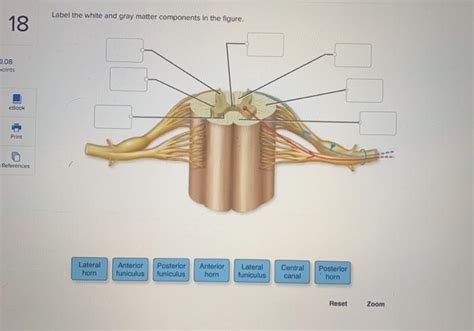
Table of Contents
Labeling the White and Gray Matter Components in the Figure: A Comprehensive Guide
Understanding the intricate architecture of the brain requires familiarity with its fundamental components: white matter and gray matter. This article provides a comprehensive guide to identifying and labeling these components within a typical brain image, exploring their distinct structures, functions, and clinical significance. We'll delve into the microscopic anatomy, macroscopic organization, and the implications of disruptions to these crucial brain regions.
Differentiating White and Gray Matter: A Macroscopic Perspective
Before we dive into labeling specific components, it's crucial to establish a clear visual distinction between white and gray matter. On a macroscopic level, the difference is readily apparent:
-
Gray matter: Appears grayish-brown in fresh tissue. It's primarily composed of neuronal cell bodies, dendrites, and axons' unmyelinated portions. It's concentrated in specific brain regions, forming a cortical sheet over the cerebrum and cerebellum, as well as deeper structures like the basal ganglia and brainstem nuclei.
-
White matter: Exhibits a white, glistening appearance due to the myelin sheaths surrounding the axons. Myelin, a fatty substance, acts as an insulator, facilitating faster nerve impulse transmission. White matter tracts connect different gray matter regions, enabling communication and coordination throughout the brain. It forms the bulk of the deep cerebral structures, the corpus callosum, and the tracts connecting the cerebrum to other brain regions.
Microscopic Anatomy: A Closer Look
The macroscopic distinction between white and gray matter reflects underlying microscopic differences:
Gray Matter Composition
At the microscopic level, gray matter is a densely packed network of:
-
Neurons: The fundamental units of the nervous system, responsible for processing and transmitting information. They have a cell body (soma), dendrites (receiving signals), and an axon (transmitting signals).
-
Glial cells: These supporting cells are crucial for neuronal function, providing structural support, insulation (myelin for oligodendrocytes), and nutrient supply. Astrocytes, oligodendrocytes, and microglia are the main types present in gray matter.
-
Synapses: The specialized junctions where communication occurs between neurons. Neurotransmitters are released here, allowing for signal transmission across the synaptic cleft.
White Matter Composition
White matter is predominantly composed of:
-
Myelinated axons: Long, slender projections of neurons covered by myelin sheaths. The myelin is produced by oligodendrocytes in the central nervous system. The myelin sheaths significantly increase the speed of nerve impulse conduction.
-
Oligodendrocytes: The glial cells responsible for producing and maintaining the myelin sheaths around axons in the CNS. A single oligodendrocyte can myelinate multiple axons.
Labeling Key White and Gray Matter Structures: A Step-by-Step Guide
Let's now consider a typical brain image (e.g., a sagittal or coronal MRI or a histological section). While the exact appearance varies depending on the imaging modality and brain region, we can identify common structures:
1. Cerebral Cortex (Gray Matter): The outermost layer of the cerebrum, responsible for higher-order cognitive functions like language, memory, and reasoning. It's highly convoluted, increasing the surface area for neuronal processing. Clearly label the frontal, parietal, temporal, and occipital lobes of the cerebral cortex.
2. Basal Ganglia (Gray Matter): Deep brain structures crucial for motor control, habit formation, and procedural learning. Key structures include the caudate nucleus, putamen, globus pallidus, and substantia nigra. These are usually visible as relatively distinct gray matter masses within the white matter of the cerebrum.
3. Thalamus (Gray Matter): A relay station for sensory information, filtering and transmitting signals to the cerebral cortex. It's located deep within the brain, superior to the midbrain.
4. Hippocampus (Gray Matter): A critical structure within the limbic system, essential for memory formation and spatial navigation. It's a C-shaped structure located within the medial temporal lobe.
5. Amygdala (Gray Matter): Another key limbic system structure, playing a vital role in processing emotions, especially fear and anxiety. It’s located near the hippocampus.
6. Corpus Callosum (White Matter): The largest white matter structure in the brain, a massive bundle of axons connecting the left and right cerebral hemispheres, facilitating interhemispheric communication. It's easily identifiable as a broad band of white matter arching across the midline of the brain.
7. Internal Capsule (White Matter): A large white matter tract carrying both ascending (sensory) and descending (motor) fibers. It lies between the thalamus and basal ganglia.
8. Cerebellar Cortex (Gray Matter): The outermost layer of the cerebellum, crucial for motor coordination, balance, and posture. It's characterized by its highly folded structure, similar to the cerebral cortex.
9. Cerebellar White Matter (White Matter): The white matter deep within the cerebellum, containing tracts connecting the cerebellar cortex to other brain regions. Its branching pattern is often described as an “arbor vitae” (tree of life).
10. Brainstem (Mix of Gray and White Matter): The lower part of the brain, connecting the cerebrum and cerebellum to the spinal cord. It contains vital centers for controlling breathing, heart rate, and consciousness. Identify the midbrain, pons, and medulla oblongata, highlighting the nuclei (gray matter) and tracts (white matter) within them.
Clinical Significance of White and Gray Matter Abnormalities
Disruptions to the structure or function of white and gray matter can have significant clinical implications:
-
Stroke: Damage to blood vessels supplying the brain can lead to ischemic (lack of blood flow) or hemorrhagic (bleeding) strokes, resulting in loss of gray matter function and potentially white matter damage.
-
Multiple Sclerosis (MS): An autoimmune disease affecting the myelin sheath in the central nervous system. This leads to demyelination and disruption of white matter tracts, causing a range of neurological symptoms.
-
Traumatic Brain Injury (TBI): Head injuries can damage both gray and white matter, resulting in cognitive, motor, and sensory deficits depending on the location and severity of the injury.
-
Neurodegenerative Diseases: Conditions like Alzheimer's disease and Parkinson's disease are associated with progressive loss of gray matter neurons and, in some cases, white matter damage.
-
Developmental Disorders: Conditions such as autism spectrum disorder and attention-deficit/hyperactivity disorder (ADHD) are often associated with structural and functional alterations in both gray and white matter.
Advanced Imaging Techniques for Detailed Visualization
Advanced neuroimaging techniques provide detailed visualizations of brain structures, enabling more precise labeling and analysis of white and gray matter:
-
Magnetic Resonance Imaging (MRI): Produces high-resolution images of brain tissues, allowing for clear differentiation between white and gray matter. Different MRI sequences (e.g., T1-weighted, T2-weighted) provide contrasting visualization of these tissues.
-
Diffusion Tensor Imaging (DTI): A type of MRI that measures the diffusion of water molecules along white matter tracts. This allows for visualization of the structural integrity and connectivity of white matter pathways.
-
Functional MRI (fMRI): Measures brain activity by detecting changes in blood flow. This allows for the identification of regions involved in specific cognitive or motor tasks, correlating gray matter activity with function.
Conclusion
The accurate labeling of white and gray matter components in brain images is crucial for understanding brain structure and function. This article provides a comprehensive overview of these fundamental components, detailing their microscopic and macroscopic characteristics and clinical relevance. Advanced imaging techniques provide further tools for detailed visualization and analysis, contributing significantly to our understanding of neuroanatomy and neurophysiology. By recognizing the differences and the interconnectivity between white and gray matter, we gain a deeper appreciation of the brain's intricate workings and the impact of various neurological conditions. Remember to always consult with a qualified medical professional for diagnosis and treatment of any neurological concerns.
Latest Posts
Latest Posts
-
Will Jill And Phil Are All Wheat Farmers
Mar 21, 2025
-
Managers Who Advocate Job Enrichment Focus On Creating Jobs With
Mar 21, 2025
-
A Patient Is Put On Medication At 20 Mg
Mar 21, 2025
-
An Account Is Said To Have A Debit Balance If
Mar 21, 2025
-
Classify The Radicals Into The Appropriate Categories
Mar 21, 2025
Related Post
Thank you for visiting our website which covers about Label The White And Gray Matter Components In The Figure . We hope the information provided has been useful to you. Feel free to contact us if you have any questions or need further assistance. See you next time and don't miss to bookmark.
