Label The Structures Of The Knee
Holbox
Mar 17, 2025 · 6 min read
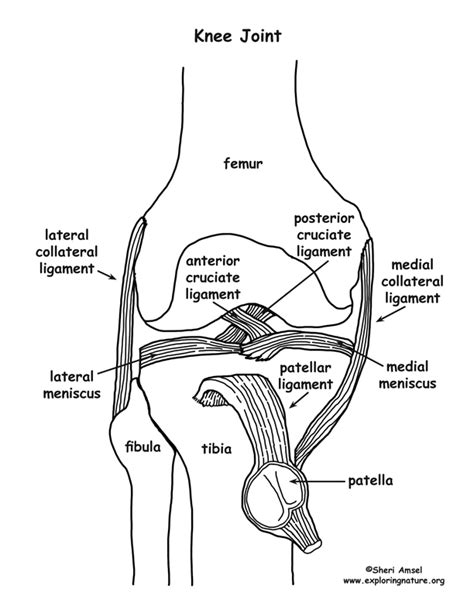
Table of Contents
Label the Structures of the Knee: A Comprehensive Guide
The knee, the largest joint in the human body, is a marvel of engineering, allowing for a wide range of motion while bearing significant weight. Understanding its intricate anatomy is crucial for anyone interested in sports medicine, physical therapy, or simply maintaining their own physical well-being. This comprehensive guide will walk you through the key structures of the knee, explaining their functions and interrelationships. We'll cover everything from bones and ligaments to cartilage and menisci, providing a detailed anatomical map of this complex joint.
The Bones of the Knee
The knee joint is formed by the articulation of three bones:
1. Femur (Thigh Bone):
The femur, the longest and strongest bone in the body, contributes its distal (lower) end to the knee joint. The distal femur features two prominent rounded condyles: the medial condyle and the lateral condyle. These condyles articulate with the tibia and patella. The smooth articular surfaces of these condyles are crucial for low-friction movement. Notice also the intercondylar fossa, a depression located between the condyles on the posterior aspect of the femur.
2. Tibia (Shin Bone):
The tibia, or shin bone, receives the weight of the body from the femur. Its proximal (upper) end features two flattened articular surfaces: the medial condyle and the lateral condyle. These condyles are slightly concave and articulate with the femoral condyles. Superior to these condyles is the tibial plateau, a relatively flat surface which provides a broad base for weight distribution. The intercondylar eminence, a prominent bony ridge between the tibial condyles, plays a crucial role in knee stability.
3. Patella (Kneecap):
The patella, a sesamoid bone (a bone embedded in a tendon), sits within the quadriceps tendon, anterior to the knee joint. It acts as a pulley, improving the efficiency of the quadriceps muscle in extending the knee. The posterior surface of the patella articulates with the patellar surface of the femur, a smooth groove on the anterior aspect of the distal femur. The patella's unique structure helps to protect the knee joint and enhance its mechanical advantage.
The Cartilage of the Knee
Cartilage plays a vital role in cushioning and protecting the knee joint, facilitating smooth movement and reducing friction.
1. Articular Cartilage:
This smooth, hyaline cartilage covers the articular surfaces of the femoral and tibial condyles, and the patella. Its resilient nature absorbs shock and allows for nearly frictionless movement. Degeneration of articular cartilage, a hallmark of osteoarthritis, leads to pain and stiffness.
2. Menisci:
The menisci are two C-shaped fibrocartilaginous discs located between the femoral and tibial condyles. The medial meniscus is larger and more C-shaped than the lateral meniscus. These menisci act as shock absorbers, distributing weight evenly across the joint, increasing stability, and improving congruency between the articulating surfaces. Tears in the menisci, often caused by twisting injuries, are a common knee injury.
The Ligaments of the Knee
Ligaments are strong fibrous tissues that connect bones, providing stability and limiting excessive movement. The knee possesses several crucial ligaments:
1. Cruciate Ligaments:
These ligaments cross each other within the knee joint, providing crucial stability against anterior-posterior displacement.
- Anterior Cruciate Ligament (ACL): Prevents the tibia from sliding forward relative to the femur. ACL tears are frequently seen in sports involving sudden changes in direction.
- Posterior Cruciate Ligament (PCL): Prevents the tibia from sliding backward relative to the femur. PCL injuries are less common than ACL injuries.
2. Collateral Ligaments:
These ligaments provide stability against medial-lateral forces.
- Medial Collateral Ligament (MCL): Located on the medial side of the knee, this ligament prevents the knee from bending inwards excessively.
- Lateral Collateral Ligament (LCL): Located on the lateral side of the knee, this ligament prevents the knee from bending outwards excessively.
The Muscles and Tendons of the Knee
Several muscles and their associated tendons contribute to the movement and stability of the knee.
1. Quadriceps Muscles:
The quadriceps femoris muscle group, consisting of the rectus femoris, vastus lateralis, vastus medialis, and vastus intermedius, is located on the anterior thigh. Its tendons converge to form the quadriceps tendon, which inserts onto the patella. The patellar tendon then connects the patella to the tibial tuberosity. The quadriceps are primarily responsible for extending the knee.
2. Hamstring Muscles:
The hamstring muscle group, located on the posterior thigh, consists of the biceps femoris, semitendinosus, and semimembranosus. These muscles flex the knee and extend the hip.
3. Other Muscles:
Several other muscles contribute to knee movement, including the popliteus muscle, which helps to unlock the knee from full extension, and the gastrocnemius muscle, a major calf muscle that contributes to knee flexion.
Bursae of the Knee
Bursae are small, fluid-filled sacs that cushion and reduce friction between structures in the knee. Several bursae surround the knee joint, including the prepatellar bursa, infrapatellar bursa, and suprapatellar bursa. Bursitis, or inflammation of the bursa, can cause pain and swelling.
Understanding Knee Injuries: A Closer Look
The complexity of the knee joint makes it susceptible to a range of injuries. Understanding the structures involved is crucial for diagnosis and treatment.
Common Knee Injuries and Their Associated Structures:
- ACL Tears: Involve damage to the anterior cruciate ligament, often resulting from sudden twisting or hyperextension.
- Meniscus Tears: Damage to the menisci, frequently due to twisting injuries, resulting in pain, swelling, and limited range of motion.
- MCL and LCL Sprains: These injuries involve stretching or tearing of the medial or lateral collateral ligaments, often caused by direct blows to the knee.
- Patellar Tendinitis (Jumper's Knee): Inflammation of the patellar tendon, commonly seen in athletes who perform repetitive jumping movements.
- Osteoarthritis: Degeneration of the articular cartilage, leading to pain, stiffness, and reduced mobility.
Understanding the specific structures involved in each injury is essential for appropriate diagnosis and treatment planning. This knowledge is also critical for injury prevention through targeted strengthening and conditioning programs.
Maintaining Knee Health: Tips and Recommendations
Maintaining knee health requires a multifaceted approach encompassing exercise, diet, and lifestyle choices.
Key Strategies for Knee Health:
- Regular Exercise: Strength training, particularly focusing on the quadriceps and hamstrings, helps stabilize the knee joint. Low-impact exercises, such as swimming and cycling, are beneficial for maintaining flexibility and range of motion.
- Proper Nutrition: A balanced diet rich in calcium, vitamin D, and other essential nutrients supports bone health.
- Maintaining a Healthy Weight: Excess weight increases stress on the knee joint, increasing the risk of injuries and osteoarthritis.
- Appropriate Footwear: Wearing supportive shoes can help to reduce stress on the knees.
- Warm-up and Cool-down: Always warm up before physical activity and cool down afterward to prepare your muscles and prevent injury.
- Proper Technique: Using correct technique during physical activities minimizes the risk of knee injuries.
- Listen to Your Body: Pay attention to any pain or discomfort in your knees and seek professional medical advice if necessary.
Conclusion
The knee joint, a marvel of biological engineering, is a complex structure composed of interconnected bones, cartilage, ligaments, muscles, and tendons. Understanding the anatomy of the knee is fundamental to appreciating its function, preventing injuries, and managing conditions that affect this vital joint. By integrating this knowledge into your exercise routines, dietary choices, and overall lifestyle, you can significantly enhance your knee health and enjoy a more active and fulfilling life. Remember that this information is for educational purposes only, and professional medical advice should always be sought for any concerns regarding your knee health.
Latest Posts
Latest Posts
-
Which Of The Following Costs Is Inventories Whehn Using
Mar 17, 2025
-
The Somatosensory Cortex Is Responsible For Processing
Mar 17, 2025
-
What Do Your Results Indicate About Cell Cycle Control
Mar 17, 2025
-
The Criteria Retailer Must Meet To Receive A Reduced Penalty
Mar 17, 2025
-
Select The Descriptions That Apply To The Thylakoid
Mar 17, 2025
Related Post
Thank you for visiting our website which covers about Label The Structures Of The Knee . We hope the information provided has been useful to you. Feel free to contact us if you have any questions or need further assistance. See you next time and don't miss to bookmark.
