Label The Longitudinal Section Of The Spinal Cord
Holbox
Mar 24, 2025 · 8 min read
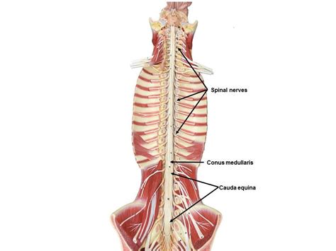
Table of Contents
- Label The Longitudinal Section Of The Spinal Cord
- Table of Contents
- Labeling the Longitudinal Section of the Spinal Cord: A Comprehensive Guide
- Key Structures of the Spinal Cord: A Preliminary Overview
- 1. Gray Matter and White Matter: The Foundation of Function
- 2. Roots and Spinal Nerves: The Connection to the Peripheral Nervous System
- 3. Meninges: Protective Layers
- 4. Conus Medullaris and Filum Terminale: The Caudal End
- 5. Cauda Equina: The "Horse's Tail"
- Step-by-Step Guide to Labeling a Longitudinal Section
- Understanding the Functional Significance of Spinal Cord Structures
- Clinical Significance: Understanding Neurological Disorders
- Latest Posts
- Latest Posts
- Related Post
Labeling the Longitudinal Section of the Spinal Cord: A Comprehensive Guide
The spinal cord, a crucial component of the central nervous system, is a fascinating structure responsible for transmitting signals between the brain and the rest of the body. Understanding its anatomy is fundamental to comprehending neurological function and dysfunction. This comprehensive guide will walk you through the process of labeling a longitudinal section of the spinal cord, highlighting key anatomical features and their significance. We'll delve into the intricate details, ensuring a thorough understanding of this vital structure.
Key Structures of the Spinal Cord: A Preliminary Overview
Before we dive into labeling, let's review the primary structures visible in a longitudinal section of the spinal cord. This foundational knowledge will make the labeling process significantly easier and more meaningful.
1. Gray Matter and White Matter: The Foundation of Function
The spinal cord is broadly divided into two distinct regions: gray matter and white matter. These areas differ significantly in their composition and function.
-
Gray Matter: This butterfly-shaped or "H"-shaped region in the center of the spinal cord is primarily composed of neuronal cell bodies, dendrites, and unmyelinated axons. It's the site of synaptic transmission and crucial for processing information. Within the gray matter, you'll find:
-
Anterior Horns (Ventral Horns): These are the larger anterior projections of the gray matter, containing motor neurons whose axons leave the spinal cord via the ventral roots to innervate skeletal muscles. Think of these as the "outgoing" pathways for motor commands.
-
Posterior Horns (Dorsal Horns): These are the smaller posterior projections, receiving sensory information from the body via the dorsal roots. These are the "incoming" pathways for sensory information.
-
Lateral Horns: Located in the thoracic and upper lumbar regions, these are smaller projections involved in the autonomic nervous system, controlling involuntary functions like heart rate and digestion.
-
-
White Matter: Surrounding the gray matter, the white matter is composed primarily of myelinated axons. Myelin, the fatty substance surrounding axons, allows for rapid signal transmission. The white matter is organized into ascending and descending tracts, facilitating communication between different levels of the spinal cord and the brain. These tracts are further divided into:
-
Ascending Tracts: These carry sensory information from the body up to the brain.
-
Descending Tracts: These carry motor commands from the brain down to the muscles.
-
2. Roots and Spinal Nerves: The Connection to the Peripheral Nervous System
The spinal cord doesn't exist in isolation. It connects to the peripheral nervous system through a series of roots and spinal nerves:
-
Dorsal Roots: These are sensory roots, carrying afferent (sensory) information into the spinal cord. Each dorsal root contains a dorsal root ganglion, a collection of sensory neuron cell bodies.
-
Ventral Roots: These are motor roots, carrying efferent (motor) information out of the spinal cord.
-
Spinal Nerves: Formed by the union of dorsal and ventral roots, these nerves carry both sensory and motor information to and from specific regions of the body.
3. Meninges: Protective Layers
The spinal cord is protected by three layers of connective tissue called meninges:
-
Dura Mater: The outermost tough, fibrous layer.
-
Arachnoid Mater: The middle layer, a delicate web-like structure. The subarachnoid space, located between the arachnoid and pia mater, contains cerebrospinal fluid (CSF).
-
Pia Mater: The innermost layer, a thin, transparent membrane that adheres directly to the spinal cord.
4. Conus Medullaris and Filum Terminale: The Caudal End
The spinal cord doesn't extend the entire length of the vertebral column. It tapers to a cone-shaped structure called the conus medullaris at approximately the L1-L2 vertebral level. From the conus medullaris, a fibrous strand called the filum terminale extends downwards, anchoring the spinal cord to the coccyx.
5. Cauda Equina: The "Horse's Tail"
Below the conus medullaris, the nerve roots extending from the lower spinal cord resemble a horse's tail, hence the term cauda equina. These nerve roots travel downwards before exiting the vertebral column at their respective levels.
Step-by-Step Guide to Labeling a Longitudinal Section
Now that we've reviewed the key structures, let's proceed to labeling a longitudinal section of the spinal cord. Imagine you have a diagram or a microscopic image in front of you. Here's a step-by-step guide:
-
Identify the Gray Matter: Begin by locating the central, butterfly-shaped gray matter. Clearly label it as "Gray Matter".
-
Differentiate Anterior and Posterior Horns: Within the gray matter, identify the larger anterior projections ("Anterior Horns" or "Ventral Horns") and the smaller posterior projections ("Posterior Horns" or "Dorsal Horns"). Note their relative sizes and positions.
-
Locate Lateral Horns (if present): If your section is from the thoracic or upper lumbar region, look for the smaller lateral projections of the gray matter and label them as "Lateral Horns".
-
Identify White Matter: Surround the gray matter, you'll find the white matter. Label this region as "White Matter".
-
Label Dorsal and Ventral Roots: Identify the dorsal and ventral roots emerging from the spinal cord. Label them clearly as "Dorsal Root" and "Ventral Root". Note the location of the "Dorsal Root Ganglion" (a swelling on the dorsal root).
-
Identify Spinal Nerve: Where the dorsal and ventral roots join, label the structure as "Spinal Nerve".
-
Label Meninges (if visible): If the meninges are depicted, label the "Dura Mater", "Arachnoid Mater", and "Pia Mater". Indicate the "Subarachnoid Space" containing CSF.
-
Locate Conus Medullaris (if applicable): If your section shows the caudal end of the spinal cord, label the tapered region as "Conus Medullaris".
-
Locate Filum Terminale (if applicable): If visible, label the fibrous extension from the conus medullaris as "Filum Terminale".
-
Label Cauda Equina (if applicable): If the section shows the nerve roots below the conus medullaris, label them as "Cauda Equina".
Understanding the Functional Significance of Spinal Cord Structures
Labeling the structures is only half the battle. Understanding their functional roles is crucial. Let's revisit the key structures and explore their functions in more detail:
Gray Matter: The gray matter is where the "action" happens. It's the site of information processing within the spinal cord.
-
Anterior Horns (Motor Neurons): These neurons initiate voluntary movements by sending signals to skeletal muscles. Damage to these neurons can lead to paralysis or weakness.
-
Posterior Horns (Sensory Neurons): These neurons receive sensory information from various receptors throughout the body, including touch, pain, temperature, and proprioception (sense of body position). Damage can result in sensory loss or disturbances.
-
Lateral Horns (Autonomic Neurons): These neurons regulate involuntary functions such as heart rate, blood pressure, digestion, and respiration. Dysfunction in these areas can cause a variety of autonomic disorders.
White Matter: The white matter serves as the communication highway, facilitating the rapid transmission of signals.
-
Ascending Tracts: These carry sensory information from the body to the brain, allowing us to perceive sensations and react to stimuli. Examples include the spinothalamic tract (pain and temperature) and the dorsal column-medial lemniscus pathway (touch and proprioception).
-
Descending Tracts: These carry motor commands from the brain to the spinal cord and muscles, enabling voluntary movement. Examples include the corticospinal tract (voluntary movement) and the reticulospinal tract (posture and balance).
Roots and Spinal Nerves: These structures form the crucial link between the central and peripheral nervous systems. Damage to these structures can result in loss of function in specific parts of the body.
Meninges: These protective layers cushion and support the spinal cord, safeguarding it from injury and infection.
Conus Medullaris, Filum Terminale, and Cauda Equina: These structures ensure the structural integrity of the spinal cord at its caudal end, facilitating the passage of nerve roots to their respective exit points.
Clinical Significance: Understanding Neurological Disorders
Understanding the anatomy of the spinal cord is vital for diagnosing and treating neurological disorders. Many conditions affect the spinal cord, including:
-
Spinal Cord Injury: Trauma can damage the spinal cord, resulting in paralysis or sensory loss depending on the location and severity of the injury.
-
Multiple Sclerosis (MS): This autoimmune disease damages the myelin sheath, impairing nerve signal transmission.
-
Amyotrophic Lateral Sclerosis (ALS): Also known as Lou Gehrig's disease, ALS is a progressive neurodegenerative disease affecting motor neurons, leading to muscle weakness and atrophy.
-
Spinal Muscular Atrophy (SMA): A genetic disorder affecting motor neurons, leading to muscle weakness and atrophy.
-
Syringomyelia: Formation of a fluid-filled cyst within the spinal cord, compressing and damaging neural tissue.
-
Tumors: Tumors within or near the spinal cord can compress and damage the neural tissue, causing various neurological symptoms.
By accurately labeling and understanding the structures of a longitudinal section of the spinal cord, healthcare professionals can better interpret diagnostic images and formulate treatment plans for these and other conditions. A solid grasp of spinal cord anatomy is therefore indispensable for anyone involved in the fields of neurology, neurosurgery, and related healthcare professions. This detailed understanding empowers accurate diagnosis, effective treatment strategies, and ultimately, improved patient outcomes.
Latest Posts
Latest Posts
-
When The Federal Reserve Conducts Open Market Operations It
Mar 29, 2025
-
The Term Permanent Current Assets Implies
Mar 29, 2025
-
A Gnat Takes Off From One End Of A Pencil
Mar 29, 2025
-
If A Management Team Wishes To Boost
Mar 29, 2025
-
Working In A Comfortable Environment With Enough Heat And Air
Mar 29, 2025
Related Post
Thank you for visiting our website which covers about Label The Longitudinal Section Of The Spinal Cord . We hope the information provided has been useful to you. Feel free to contact us if you have any questions or need further assistance. See you next time and don't miss to bookmark.
