Label The Drawing Of The Nephron Using The Key Letters
Holbox
Mar 27, 2025 · 6 min read
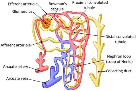
Table of Contents
- Label The Drawing Of The Nephron Using The Key Letters
- Table of Contents
- Label the Drawing of the Nephron Using the Key Letters: A Comprehensive Guide
- Key Structures of the Nephron: A Detailed Breakdown
- 1. The Renal Corpuscle: The Filtration Unit
- 2. The Renal Tubule: Reabsorption and Secretion
- Labeling Your Nephron Diagram: A Step-by-Step Guide
- Understanding Nephron Function: Clinical Significance
- Beyond the Basics: Advanced Nephron Anatomy and Physiology
- Latest Posts
- Latest Posts
- Related Post
Label the Drawing of the Nephron Using the Key Letters: A Comprehensive Guide
The nephron, the functional unit of the kidney, is a complex structure responsible for filtering blood and producing urine. Understanding its intricate anatomy is crucial for comprehending renal physiology and various kidney-related diseases. This comprehensive guide will walk you through labeling a nephron diagram, explaining the function of each component in detail. We'll cover everything from the glomerulus to the collecting duct, ensuring you master the anatomy of this vital organ.
Key Structures of the Nephron: A Detailed Breakdown
Before we delve into labeling, let's review the key structures of the nephron. A typical nephron consists of two main parts: the renal corpuscle and the renal tubule.
1. The Renal Corpuscle: The Filtration Unit
The renal corpuscle, located in the cortex of the kidney, is responsible for the initial filtration of blood. It comprises two key structures:
-
Glomerulus (A): A network of capillaries where blood is initially filtered. The high pressure within the glomerulus forces fluid and small solutes across the capillary walls. This process is crucial for the removal of waste products from the blood. Remember: The glomerulus is surrounded by Bowman's capsule.
-
Bowman's Capsule (B): A double-walled cup-shaped structure that surrounds the glomerulus. The filtrate, a mixture of water, small solutes, and some proteins, passes from the glomerulus into the Bowman's space within Bowman's capsule. The inner layer of Bowman's capsule is composed of specialized cells called podocytes, crucial for selective filtration. Important: The filtration membrane, composed of fenestrated endothelium, basement membrane, and podocyte foot processes, prevents the passage of large molecules like blood cells and plasma proteins.
2. The Renal Tubule: Reabsorption and Secretion
The renal tubule, a long, convoluted tube, extends from Bowman's capsule. It's responsible for the fine-tuning of the filtrate's composition through reabsorption and secretion. The renal tubule is divided into several segments:
-
Proximal Convoluted Tubule (PCT) (C): This segment is characterized by its extensive microvilli, which increase the surface area for reabsorption. The PCT reabsorbs the majority of essential substances from the filtrate, including glucose, amino acids, water, sodium, potassium, chloride, and bicarbonate ions. It also secretes certain substances like hydrogen and ammonium ions. Key Point: The PCT is crucial for maintaining the body's fluid and electrolyte balance.
-
Loop of Henle (D): This U-shaped structure extends from the PCT into the medulla of the kidney. It plays a critical role in concentrating the urine. The descending limb is highly permeable to water but impermeable to solutes, while the ascending limb is impermeable to water but actively transports sodium and chloride ions out of the filtrate. Understanding the Countercurrent Mechanism: The Loop of Henle's unique structure allows for the establishment of a concentration gradient in the medulla, which facilitates water reabsorption from the collecting duct.
-
Distal Convoluted Tubule (DCT) (E): Located in the cortex, the DCT plays a crucial role in fine-tuning the electrolyte balance. It reabsorbs sodium and calcium ions and secretes potassium and hydrogen ions. The DCT is also influenced by hormones such as aldosterone and parathyroid hormone, which regulate its function. Note: The DCT is responsible for maintaining acid-base balance.
-
Collecting Duct (F): The collecting duct receives filtrate from several nephrons. Its primary role is to regulate the final concentration of urine. The permeability of the collecting duct to water is regulated by antidiuretic hormone (ADH). In the presence of ADH, the collecting duct becomes highly permeable to water, allowing for increased water reabsorption and the production of concentrated urine. In the absence of ADH, the collecting duct is less permeable to water, resulting in dilute urine. Crucial Role: The collecting duct contributes significantly to the body's overall fluid balance and osmoregulation.
Labeling Your Nephron Diagram: A Step-by-Step Guide
Now, let's put this knowledge into practice. To accurately label your nephron diagram, follow these steps:
-
Identify the Renal Corpuscle: Locate the glomerulus (A) and Bowman's capsule (B). The glomerulus should appear as a network of capillaries nestled within the Bowman's capsule.
-
Trace the Renal Tubule: Follow the pathway of the renal tubule, starting from Bowman's capsule. Identify the proximal convoluted tubule (PCT) (C), which is characterized by its convoluted shape and densely packed cells.
-
Locate the Loop of Henle: Identify the loop of Henle (D), recognizing its characteristic U-shaped structure extending into the medulla. Differentiate between the descending and ascending limbs.
-
Find the Distal Convoluted Tubule: Locate the distal convoluted tubule (DCT) (E), which is also characterized by its convoluted shape, but typically less tightly packed than the PCT.
-
Identify the Collecting Duct: Find the collecting duct (F), which receives filtrate from multiple nephrons and extends towards the renal pelvis.
-
Label Accurately: Use the key letters (A-F) to clearly label each structure on your diagram. Ensure the labels are neat, clear, and easily readable.
-
Consider Adding Additional Details: Depending on the complexity of your diagram, you may consider labeling additional structures like the juxtaglomerular apparatus (which regulates blood pressure and glomerular filtration rate), peritubular capillaries, and vasa recta.
Understanding Nephron Function: Clinical Significance
A thorough understanding of nephron function is vital for grasping various renal diseases and conditions. Here are some examples:
-
Glomerulonephritis: Inflammation of the glomerulus, often caused by autoimmune disorders or infections, can impair filtration and lead to proteinuria (protein in the urine) and hematuria (blood in the urine).
-
Acute Kidney Injury (AKI): Sudden decline in kidney function, often caused by dehydration, infections, or certain medications. AKI can lead to fluid and electrolyte imbalances and accumulation of waste products in the blood.
-
Chronic Kidney Disease (CKD): Progressive loss of kidney function over time, often caused by diabetes, hypertension, or glomerulonephritis. CKD can lead to a range of complications, including cardiovascular disease, anemia, and bone disease.
-
Diabetes Insipidus: A condition characterized by the inability to concentrate urine due to insufficient ADH production or action. This leads to excessive urination and dehydration.
Beyond the Basics: Advanced Nephron Anatomy and Physiology
While this guide focuses on the basic structures and functions of the nephron, further exploration can reveal greater complexity. For instance:
-
Juxtaglomerular Apparatus (JGA): This specialized structure, located at the junction of the DCT and the afferent arteriole, plays a critical role in regulating blood pressure and glomerular filtration rate through the renin-angiotensin-aldosterone system (RAAS).
-
Peritubular Capillaries and Vasa Recta: These capillary networks surrounding the renal tubules facilitate reabsorption and secretion. The vasa recta, specialized capillaries associated with the Loop of Henle, are crucial for maintaining the medullary concentration gradient.
-
Transport Mechanisms: The nephron utilizes various transport mechanisms, including active transport, passive transport, and facilitated diffusion, to move substances across its membranes. Understanding these mechanisms is essential for comprehending the precise control of fluid and electrolyte balance.
-
Hormonal Regulation: Several hormones, including ADH, aldosterone, and parathyroid hormone, play crucial roles in regulating nephron function. These hormones influence the reabsorption and secretion of various substances, contributing to the precise control of fluid and electrolyte balance.
By mastering the labeling of a nephron diagram and understanding the intricate functions of its components, you will gain a solid foundation in renal physiology. This knowledge is not only crucial for medical professionals but also beneficial for anyone interested in understanding the complex workings of the human body. Remember to practice labeling multiple diagrams, and consult various resources to solidify your understanding of this fascinating and vital organ system. The more you practice, the more confident and proficient you'll become in your understanding of the nephron.
Latest Posts
Latest Posts
-
Luna Mae Is 12 Years Old
Mar 31, 2025
-
Angus Works As A Dairy Farmer
Mar 31, 2025
-
All Competitive Markets Involve Which Of The Following
Mar 31, 2025
-
Write An Equation For The Function Graphed Below
Mar 31, 2025
-
A Carnot Refrigerator Absorbs Heat From A Space At 15
Mar 31, 2025
Related Post
Thank you for visiting our website which covers about Label The Drawing Of The Nephron Using The Key Letters . We hope the information provided has been useful to you. Feel free to contact us if you have any questions or need further assistance. See you next time and don't miss to bookmark.
