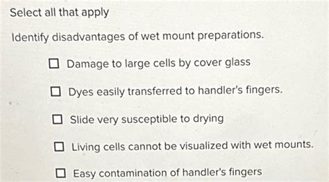
Identify Disadvantages Of Wet Mount Preparations.
Holbox
Mar 29, 2025 · 5 min read

Table of Contents
- Identify Disadvantages Of Wet Mount Preparations.
- Table of Contents
- Identifying the Disadvantages of Wet Mount Preparations
- Limitations in Specimen Visualization
- 1. Movement and Distortion of Specimens:
- 2. Limited Resolution and Contrast:
- 3. Drying Artifacts:
- 4. Obscured Details by Debris and Bubbles:
- Challenges in Specimen Preservation and Handling
- 5. Short-Term Observation Only:
- 6. Risk of Contamination:
- 7. Difficulty in Staining:
- 8. Limited Applicability to Thick Specimens:
- Comparison with Alternative Techniques
- 9. Superiority of Permanent Mounts:
- 10. Advanced Microscopic Techniques:
- Conclusion: Choosing the Right Technique
- Latest Posts
- Latest Posts
- Related Post
Identifying the Disadvantages of Wet Mount Preparations
Wet mount preparations, a simple and widely used technique in microscopy, offer a quick and easy way to visualize live specimens. However, this seemingly straightforward method comes with a number of significant disadvantages that limit its applicability and can affect the accuracy and reliability of observations. Understanding these drawbacks is crucial for choosing the appropriate microscopy technique and interpreting results accurately. This comprehensive article explores the various limitations of wet mount preparations, providing a detailed overview for researchers, students, and anyone working with microscopy.
Limitations in Specimen Visualization
One of the most significant disadvantages of wet mount preparations lies in the inherent limitations they impose on specimen visualization. These limitations can significantly impact the quality and interpretability of microscopic observations.
1. Movement and Distortion of Specimens:
Perhaps the most frustrating aspect of wet mount preparations is the uncontrolled movement of live specimens. Bacteria, protozoa, and other microorganisms can move rapidly, making it extremely difficult to focus and observe their structures clearly. This constant motion blurs the image, hindering detailed observation and accurate measurements. Moreover, the pressure of the coverslip can distort the specimen's shape, leading to inaccurate morphological assessments. This is particularly problematic when attempting to identify specific microorganisms based on their size and shape.
2. Limited Resolution and Contrast:
Wet mount preparations often suffer from low resolution and poor contrast. The aqueous medium surrounding the specimen doesn't provide optimal optical properties, leading to blurry images with reduced detail. This can be particularly challenging when dealing with small or transparent specimens where fine structures are difficult to distinguish. The lack of contrast further complicates visualization, making it difficult to differentiate between the specimen and the background. Advanced microscopy techniques, such as phase-contrast or differential interference contrast microscopy, can partially mitigate these issues, but they are often not practical for routine wet mount preparations.
3. Drying Artifacts:
The aqueous medium used in wet mount preparations is prone to evaporation, particularly during prolonged observation. As the liquid evaporates, the specimen can become desiccated, leading to shrinkage, distortion, and the formation of drying artifacts. These artifacts can alter the appearance of the specimen, making it difficult to interpret its true morphology and potentially leading to misidentification. Employing techniques like using a sealant around the edges of the coverslip can help to slow down evaporation, but it cannot completely eliminate this issue.
4. Obscured Details by Debris and Bubbles:
The preparation process itself can introduce artifacts that interfere with observation. Dust particles, debris from the slide or coverslip, and air bubbles trapped between the coverslip and the specimen can obscure fine structures and complicate visualization. These artifacts can be particularly problematic when working with small specimens or when attempting to observe delicate structures. Careful preparation techniques, including cleaning slides and coverslips thoroughly and employing proper mounting techniques, can help minimize this issue, but it cannot be eliminated entirely.
Challenges in Specimen Preservation and Handling
Beyond issues with visualization, wet mounts also present challenges in specimen preservation and handling, further limiting their utility in various applications.
5. Short-Term Observation Only:
Wet mount preparations are inherently unsuitable for long-term observation. The specimens are typically alive and metabolically active, and their condition can change rapidly over time. Changes in pH, nutrient depletion, and the accumulation of metabolic waste products can alter the specimen's appearance and behavior, impacting the accuracy of observations. This makes them unsuitable for time-lapse studies or prolonged observation of cellular processes.
6. Risk of Contamination:
The open nature of wet mount preparations makes them susceptible to contamination. Exposure to airborne microorganisms can introduce contaminants, altering the composition of the specimen and potentially compromising the accuracy of the results. This risk is especially high in environments with high microbial loads or when working with sensitive specimens. Using sterile techniques during preparation and minimizing exposure to the environment can help reduce contamination risk, but it cannot be eliminated completely.
7. Difficulty in Staining:
While some simple staining techniques can be applied to wet mount preparations, more complex or specialized staining procedures are often incompatible. The aqueous medium can interfere with the staining process, preventing the stain from penetrating the specimen effectively. This limitation restricts the ability to highlight specific cellular structures or components, limiting the scope of detailed analysis.
8. Limited Applicability to Thick Specimens:
Wet mount preparations are best suited for observing thin specimens. Thick specimens often result in an uneven distribution of the mounting medium, making it difficult to obtain a clear and focused image. Light scattering and other optical artifacts can further complicate observation, rendering detailed analysis impossible. Sectioning or other sample preparation techniques are necessary for examining thick specimens effectively.
Comparison with Alternative Techniques
Understanding the disadvantages of wet mount preparations requires comparing them with alternative microscopy techniques that address some of these limitations.
9. Superiority of Permanent Mounts:
Permanent mounts, created by embedding specimens in a resin or other mounting medium, offer several advantages over wet mounts. They prevent specimen movement, provide better contrast and resolution, and allow for long-term storage and repeated observation without concerns about specimen degradation or drying artifacts. However, this method requires additional time and specialized equipment and is not suitable for observing live specimens.
10. Advanced Microscopic Techniques:
Techniques like phase-contrast microscopy, differential interference contrast microscopy, and fluorescence microscopy offer enhanced contrast and resolution, overcoming some of the limitations of wet mounts. However, these techniques require specialized equipment and expertise, making them less accessible than the simple wet mount preparation.
Conclusion: Choosing the Right Technique
While wet mount preparations offer a simple and quick method for observing live specimens, their limitations must be carefully considered when choosing an appropriate microscopy technique. The choice between a wet mount and an alternative method depends on several factors, including the type of specimen, the desired level of detail, the duration of observation, and the available resources. Understanding these disadvantages allows researchers to make informed decisions, ensuring that the chosen method provides reliable and accurate results. The limitations discussed above highlight the importance of choosing a preparation technique that aligns with specific research goals and ensures that observations are accurate, repeatable, and meaningful. Considering these factors is crucial for optimal results in microscopic analysis.
Latest Posts
Latest Posts
-
Problems In Balance May Follow Trauma To Which Nerve
Apr 02, 2025
-
Another Term For The All Channel Communication Network Is
Apr 02, 2025
-
The Controllable Variance Is So Called Because It
Apr 02, 2025
-
The Graph Of The Relation S Is Shown Below
Apr 02, 2025
-
Which One Of The Following Is A Working Capital Decision
Apr 02, 2025
Related Post
Thank you for visiting our website which covers about Identify Disadvantages Of Wet Mount Preparations. . We hope the information provided has been useful to you. Feel free to contact us if you have any questions or need further assistance. See you next time and don't miss to bookmark.
