Correctly Label The Following Anatomical Features Of The Eye
Holbox
Mar 24, 2025 · 6 min read
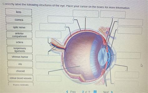
Table of Contents
- Correctly Label The Following Anatomical Features Of The Eye
- Table of Contents
- Correctly Labeling the Anatomical Features of the Eye: A Comprehensive Guide
- The Outer Layer: Protection and Light Focusing
- 1. Cornea: The Transparent Shield
- 2. Sclera: The Protective White
- 3. Conjunctiva: The Mucous Membrane Lining
- The Middle Layer: The Vascular and Iris
- 4. Choroid: The Blood Supply Network
- 5. Ciliary Body: Accommodation and Aqueous Humor Production
- 6. Iris: The Color and Aperture Control
- The Inner Layer: The Retina and Photoreception
- 7. Retina: The Light-Sensitive Layer
- 8. Macula: The Central Vision Hub
- 9. Optic Disc (Blind Spot): Where the Optic Nerve Exits
- 10. Optic Nerve: The Information Highway
- The Lens: Focusing Light onto the Retina
- 11. Lens: Focusing Powerhouse
- The Aqueous and Vitreous Humors: Maintaining Eye Shape and Clarity
- 12. Aqueous Humor: The Anterior Chamber Fluid
- 13. Vitreous Humor: The Posterior Chamber Gel
- Understanding Eye Anatomy: Implications for Eye Health
- Beyond the Basics: Further Exploration of the Eye's Complexity
- Latest Posts
- Latest Posts
- Related Post
Correctly Labeling the Anatomical Features of the Eye: A Comprehensive Guide
The human eye, a marvel of biological engineering, is responsible for our sense of sight. Understanding its intricate structure is crucial for appreciating its function and diagnosing potential issues. This comprehensive guide provides a detailed explanation of the eye's anatomical features, ensuring you can correctly label each component. We'll explore the structures from the outermost layers to the innermost, focusing on their roles in vision and overall eye health.
The Outer Layer: Protection and Light Focusing
The outermost layer of the eye provides protection and plays a key role in focusing light onto the retina. This layer consists of three main components:
1. Cornea: The Transparent Shield
The cornea is the transparent, dome-shaped outer layer covering the front of the eye. It's responsible for refracting (bending) light as it enters the eye, contributing significantly to the eye's focusing power. Its clarity is vital for sharp vision; any clouding can severely impair sight. The cornea is highly sensitive to touch, triggering the blink reflex to protect it from potential harm.
2. Sclera: The Protective White
The sclera is the tough, white, fibrous outer layer that forms the majority of the eyeball's surface. It provides structural support and protection for the delicate inner structures. The visible white part of the eye is the sclera. Tiny blood vessels within the sclera provide nourishment to the eye tissues.
3. Conjunctiva: The Mucous Membrane Lining
The conjunctiva is a thin, transparent mucous membrane that lines the inside of the eyelids and covers the sclera. It helps to lubricate the eye and protect it from infection. Inflammation of the conjunctiva, known as conjunctivitis (pink eye), is a common condition characterized by redness, itching, and discharge.
The Middle Layer: The Vascular and Iris
The middle layer of the eye, also known as the uvea, is rich in blood vessels and plays a crucial role in regulating the amount of light entering the eye. Key structures within this layer include:
4. Choroid: The Blood Supply Network
The choroid is a highly vascularized layer located between the sclera and the retina. Its primary function is to supply blood and nutrients to the outer layers of the retina. Its rich blood supply is essential for maintaining the retina's metabolic activity and visual function.
5. Ciliary Body: Accommodation and Aqueous Humor Production
The ciliary body is a ring-shaped structure located behind the iris. It contains the ciliary muscles, which control the shape of the lens, enabling the eye to focus on objects at different distances (a process called accommodation). The ciliary body also produces aqueous humor, the clear fluid that fills the anterior chamber of the eye.
6. Iris: The Color and Aperture Control
The iris is the colored part of the eye, responsible for controlling the amount of light that enters the pupil. It contains two sets of muscles: the circular muscles (constrictor pupillae) that constrict the pupil in bright light, and the radial muscles (dilator pupillae) that dilate the pupil in dim light. The color of the iris is determined by the amount and distribution of melanin pigment.
The Inner Layer: The Retina and Photoreception
The innermost layer of the eye is the retina, the light-sensitive layer that converts light into electrical signals that the brain can interpret as images. The retina is incredibly complex, containing millions of specialized cells:
7. Retina: The Light-Sensitive Layer
The retina is a thin, delicate layer of tissue lining the back of the eye. It contains photoreceptor cells called rods and cones, which are responsible for detecting light. Rods are highly sensitive to light and responsible for vision in low-light conditions. Cones are responsible for color vision and visual acuity in bright light.
8. Macula: The Central Vision Hub
The macula is a small, specialized area in the central retina responsible for sharp, central vision. Within the macula is the fovea, a tiny pit containing a high concentration of cones, providing the highest visual acuity. Damage to the macula, as in macular degeneration, significantly impairs central vision.
9. Optic Disc (Blind Spot): Where the Optic Nerve Exits
The optic disc is the area where the optic nerve exits the eye. It lacks photoreceptor cells, resulting in a blind spot in our visual field. Our brain typically compensates for this blind spot, so we're usually unaware of its presence.
10. Optic Nerve: The Information Highway
The optic nerve is a bundle of nerve fibers that carries visual information from the retina to the brain. It transmits the electrical signals generated by the photoreceptor cells, allowing us to perceive images.
The Lens: Focusing Light onto the Retina
Located behind the iris and pupil, the lens is a transparent, biconvex structure that plays a crucial role in focusing light onto the retina. Its flexibility allows it to adjust its shape to focus on objects at various distances:
11. Lens: Focusing Powerhouse
The lens is made of specialized protein fibers arranged in a layered structure. Its shape is controlled by the ciliary muscles. As we age, the lens loses its flexibility, leading to a condition called presbyopia, which makes it difficult to focus on near objects. Cataracts, a clouding of the lens, also affect focusing ability.
The Aqueous and Vitreous Humors: Maintaining Eye Shape and Clarity
The eye's internal chambers are filled with fluids that maintain its shape and contribute to its optical properties:
12. Aqueous Humor: The Anterior Chamber Fluid
Aqueous humor is a clear, watery fluid that fills the anterior chamber (the space between the cornea and the lens). It provides nutrients to the cornea and lens and helps maintain the intraocular pressure (pressure inside the eye). Problems with aqueous humor production or drainage can lead to glaucoma.
13. Vitreous Humor: The Posterior Chamber Gel
Vitreous humor is a clear, gel-like substance that fills the posterior chamber (the space between the lens and the retina). It helps maintain the shape of the eyeball and contributes to the eye's optical properties. Floaters, tiny specks that appear in the vision, are often caused by changes in the vitreous humor.
Understanding Eye Anatomy: Implications for Eye Health
A thorough understanding of the eye's anatomical features is paramount for maintaining good eye health. Knowing the function of each structure allows us to better appreciate the potential impact of eye diseases and conditions. Regular eye exams are crucial for early detection and treatment of issues such as glaucoma, cataracts, macular degeneration, and diabetic retinopathy. Early intervention often significantly improves visual outcomes and overall quality of life.
Beyond the Basics: Further Exploration of the Eye's Complexity
This detailed guide provides a comprehensive overview of the eye's key anatomical features. However, the eye's intricate structure extends beyond these fundamental components. Further exploration can delve into the neuroanatomy of vision, the biochemistry of photoreception, and the sophisticated neural pathways involved in processing visual information. Understanding these advanced aspects requires further study in ophthalmology and neuroscience.
This detailed guide provides a robust foundation for correctly labeling and understanding the anatomical structures of the human eye. Remember, this is a complex organ with interconnected parts. Regular eye care and a solid understanding of its anatomy are vital for preserving your vision.
Latest Posts
Latest Posts
-
The Belmont Principle Of Beneficence Requires That
Mar 28, 2025
-
Robin Would Like To Shoot An Orange
Mar 28, 2025
-
Who Are The Culture Ambassador At Infosys
Mar 28, 2025
-
Written Assignment 7 Dilations And Symmetry
Mar 28, 2025
-
Populating An Array With A For Loop
Mar 28, 2025
Related Post
Thank you for visiting our website which covers about Correctly Label The Following Anatomical Features Of The Eye . We hope the information provided has been useful to you. Feel free to contact us if you have any questions or need further assistance. See you next time and don't miss to bookmark.
