Correctly Label The Following Anatomical Features Of A Neuron
Holbox
Mar 24, 2025 · 6 min read
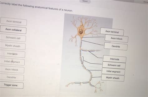
Table of Contents
- Correctly Label The Following Anatomical Features Of A Neuron
- Table of Contents
- Correctly Labeling the Anatomical Features of a Neuron: A Comprehensive Guide
- The Neuron: A Cellular Overview
- Key Anatomical Features and Their Functions
- 1. Soma (Cell Body)
- 2. Dendrites
- 3. Axon
- 4. Axon Hillock
- 5. Myelin Sheath
- 6. Nodes of Ranvier
- 7. Axon Terminals (Terminal Buttons or Synaptic Boutons)
- 8. Synapse
- 9. Synaptic Vesicles
- 10. Neurotransmitters
- Clinical Significance of Understanding Neuronal Anatomy
- Conclusion: Mastering Neuronal Anatomy
- Latest Posts
- Latest Posts
- Related Post
Correctly Labeling the Anatomical Features of a Neuron: A Comprehensive Guide
Understanding the intricacies of a neuron, the fundamental unit of the nervous system, is crucial for comprehending how our brains and bodies function. This guide provides a detailed exploration of neuronal anatomy, equipping you with the knowledge to correctly label its various components. We'll delve into the structure, function, and significance of each part, ensuring a thorough understanding of this fascinating cell.
The Neuron: A Cellular Overview
Neurons, also known as nerve cells, are specialized cells designed for the rapid transmission of electrical and chemical signals throughout the body. This communication is vital for everything from basic reflexes to complex cognitive processes. Their unique structure directly reflects their function. Unlike many other cells, neurons are generally non-replicating (though some exceptions exist in certain brain regions), meaning they don't readily undergo cell division. This contributes to the permanent nature of many neurological conditions.
Key Anatomical Features and Their Functions
Let's explore the essential parts of a neuron, focusing on their specific roles in signal transmission and processing:
1. Soma (Cell Body)
The soma, also known as the perikaryon, is the neuron's central hub. It contains the nucleus, which houses the neuron's genetic material (DNA), and other essential organelles, such as mitochondria (for energy production), ribosomes (for protein synthesis), and the endoplasmic reticulum (involved in protein folding and transport). The soma is responsible for maintaining the neuron's overall health and function. Damage to the soma is typically irreversible and leads to neuronal death.
2. Dendrites
Dendrites are branched extensions emanating from the soma. They act as the primary receivers of signals from other neurons. Their extensive branching increases the surface area available for receiving these incoming signals, known as synaptic inputs. The structure of dendrites, including their spines (small protrusions on the dendrites), plays a crucial role in synaptic plasticity, the ability of synapses to strengthen or weaken over time, a process fundamental to learning and memory. The more complex the dendritic arborization (branching pattern), the more synaptic inputs a neuron can receive.
3. Axon
The axon is a long, slender projection extending from the soma. It serves as the neuron's primary transmitter of signals. Unlike dendrites, which receive signals, the axon conducts signals away from the soma towards other neurons, muscles, or glands. The axon's length can vary significantly, ranging from a few micrometers to over a meter in length (for example, those extending from the spinal cord to the toes). The axon is often covered by a myelin sheath, a fatty insulating layer that significantly speeds up signal transmission.
4. Axon Hillock
The axon hillock is a specialized region of the neuron where the axon originates from the soma. This area plays a critical role in integrating the incoming signals from the dendrites. It acts as a decision-making point, determining whether the summed input signals are strong enough to trigger the generation of an action potential (a rapid electrical signal that travels down the axon). The axon hillock has a high concentration of voltage-gated ion channels, essential for the initiation of action potentials. The axon hillock is considered the neuron's trigger zone.
5. Myelin Sheath
The myelin sheath, mentioned previously, is a fatty insulating layer that surrounds many axons. It is formed by specialized glial cells: oligodendrocytes in the central nervous system (brain and spinal cord) and Schwann cells in the peripheral nervous system. The myelin sheath speeds up the conduction of action potentials through a process called saltatory conduction, where the signal "jumps" between the gaps in the myelin sheath, known as Nodes of Ranvier.
6. Nodes of Ranvier
Nodes of Ranvier are the gaps between segments of the myelin sheath along the axon. They are rich in voltage-gated ion channels, crucial for the regeneration of the action potential during saltatory conduction. These nodes play a vital role in ensuring the rapid and efficient transmission of the signal down the axon. Damage to the myelin sheath, as seen in diseases like multiple sclerosis, can severely impair signal transmission.
7. Axon Terminals (Terminal Buttons or Synaptic Boutons)
Axon terminals, also called terminal buttons or synaptic boutons, are the endings of the axon. These structures form synapses, specialized junctions with other neurons or target cells (muscles, glands). At the synapse, the neuron releases neurotransmitters, chemical messengers that transmit the signal to the next cell. The axon terminal contains vesicles filled with these neurotransmitters, which are released into the synaptic cleft upon arrival of the action potential.
8. Synapse
The synapse is the point of communication between two neurons or a neuron and its target cell. It comprises the presynaptic terminal (axon terminal), the synaptic cleft (the space between the two cells), and the postsynaptic membrane (the membrane of the receiving cell). The release of neurotransmitters across the synaptic cleft triggers changes in the postsynaptic cell, either exciting or inhibiting it, depending on the type of neurotransmitter and the receptor it binds to. Synaptic transmission is a complex process involving many molecules and pathways, crucial for neural communication.
9. Synaptic Vesicles
Synaptic vesicles are small sacs located within the axon terminals. They store and release neurotransmitters into the synaptic cleft. The arrival of an action potential triggers the fusion of these vesicles with the presynaptic membrane, releasing their contents. The amount and frequency of neurotransmitter release are tightly regulated and crucial for efficient signal transmission.
10. Neurotransmitters
Neurotransmitters are chemical messengers that transmit signals across the synapse from one neuron to another or to a target cell. Many different types of neurotransmitters exist, each with its own specific effects. Examples include acetylcholine, dopamine, serotonin, and glutamate. Neurotransmitters bind to specific receptors on the postsynaptic membrane, triggering a variety of cellular responses, contributing to the complex workings of the nervous system.
Clinical Significance of Understanding Neuronal Anatomy
A thorough understanding of neuronal anatomy is crucial in various medical fields. For instance, neurologists rely heavily on this knowledge to diagnose and treat neurological disorders. Damage to specific parts of the neuron can have profound consequences, leading to a wide range of symptoms. Examples include:
- Multiple sclerosis: Damage to the myelin sheath impairs signal transmission, causing a variety of neurological symptoms.
- Stroke: Disruption of blood flow to the brain can cause neuronal death and severe neurological deficits.
- Alzheimer's disease: Neuronal degeneration and synapse loss contribute to cognitive decline.
- Parkinson's disease: Loss of dopamine-producing neurons in a specific brain region leads to motor impairments.
By understanding the precise roles of each neuronal component, clinicians can better understand the mechanisms underlying neurological diseases and develop more effective treatments. Furthermore, ongoing research into neuronal structure and function continues to reveal new insights into the workings of the brain and the potential for developing novel therapeutic strategies for neurological disorders.
Conclusion: Mastering Neuronal Anatomy
Correctly labeling the anatomical features of a neuron is a fundamental step towards understanding the complexities of the nervous system. From the soma's central role in maintaining the neuron's health to the synapse's crucial role in intercellular communication, each component plays a vital role in the processing and transmission of information within our bodies. This detailed guide has provided a comprehensive overview of these components, their functions, and their clinical significance. Further exploration of neuroscience literature will provide an even deeper understanding of this fascinating subject. Remember, a strong grasp of neuronal anatomy serves as a critical foundation for advanced studies in neuroscience and related medical fields.
Latest Posts
Latest Posts
-
Which Data Type Can Only Be Classified As Text
Mar 27, 2025
-
Intellectual Property Protection And Social Complexity Are Examples Of
Mar 27, 2025
-
At Equilibrium Producer Surplus Is Represented By The Area
Mar 27, 2025
-
If The Four Firm Concentration Ratio For Industry X Is 80
Mar 27, 2025
-
Sam Is An It Manager A New Process
Mar 27, 2025
Related Post
Thank you for visiting our website which covers about Correctly Label The Following Anatomical Features Of A Neuron . We hope the information provided has been useful to you. Feel free to contact us if you have any questions or need further assistance. See you next time and don't miss to bookmark.
