Which Event Has To Precede All Others During Endochondral Ossification
Holbox
Mar 21, 2025 · 7 min read
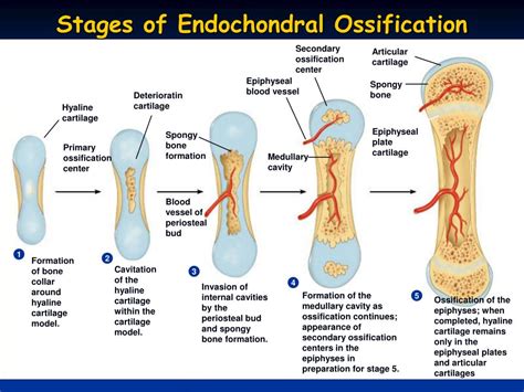
Table of Contents
- Which Event Has To Precede All Others During Endochondral Ossification
- Table of Contents
- Which Event Has to Precede All Others During Endochondral Ossification?
- The Primacy of the Cartilage Model: The Foundation of Bone Development
- The Stages Leading to Cartilage Model Formation: A Recap
- The Subsequent Stages of Endochondral Ossification: Dependent on the Cartilage Template
- The Critical Role of Signaling Pathways: Ensuring the Order of Events
- Clinical Significance: Consequences of Disruptions in Endochondral Ossification
- Conclusion: The Irreplaceable Role of the Cartilage Model
- Latest Posts
- Latest Posts
- Related Post
Which Event Has to Precede All Others During Endochondral Ossification?
Endochondral ossification, the process by which most bones in the body are formed, is a complex and precisely orchestrated sequence of events. Understanding the order of these events is crucial to grasping the intricacies of skeletal development and the potential consequences of disruptions in this process. The question, "Which event has to precede all others during endochondral ossification?" points to a fundamental step that initiates the entire cascade of cellular and molecular interactions. The answer, unequivocally, is the formation of the cartilage model.
The Primacy of the Cartilage Model: The Foundation of Bone Development
Before any bone tissue can be formed through endochondral ossification, a meticulously shaped scaffold of hyaline cartilage must exist. This cartilage model, also known as the cartilage anlage, serves as the template upon which bone will be built. Its precise shape dictates the future form of the bone. This initial stage is not merely a precursor; it's the absolute prerequisite. Without the cartilage model, the subsequent stages of endochondral ossification are impossible.
The Stages Leading to Cartilage Model Formation: A Recap
While the cartilage model's formation is the crucial initiating event, let's briefly examine the stages preceding its full development to fully appreciate its centrality:
-
Mesenchymal Condensation: This is the very first step. Mesenchymal cells, undifferentiated connective tissue cells, aggregate at specific locations determined by genetic and signaling pathways. These cells, highly responsive to various growth factors and morphogens, clump together forming a dense mass. This condensation is not random; it is guided by intricate signaling networks ensuring the proper location and shape of the future bone.
-
Differentiation into Chondrocytes: Within the mesenchymal condensation, cells undergo differentiation, transforming into chondrocytes – the specialized cells that produce and maintain the cartilage matrix. This differentiation involves the activation of specific genes and the expression of cartilage-specific proteins such as collagen type II, aggrecan, and other extracellular matrix components. The expression of these proteins is critical; their absence would prevent the formation of the cartilage matrix, thus halting the entire process.
-
Cartilage Matrix Secretion: The newly differentiated chondrocytes begin to secrete the extracellular matrix (ECM) of hyaline cartilage. This matrix consists of collagen fibers, proteoglycans, and other molecules that provide structural support and contribute to the cartilage's unique properties. The secretion of the matrix expands the cartilage model, shaping it into a recognizable form reflecting the future bone. The matrix itself is a highly dynamic environment, undergoing constant remodeling as the chondrocytes continue their activity.
-
Formation of the Complete Cartilage Model: The process of chondrocyte differentiation and matrix secretion continues until a complete cartilage model, mirroring the shape of the future bone, is formed. This model is not simply a homogeneous mass; it features distinct zones reflecting the varying stages of chondrocyte differentiation and matrix composition. These zones (reserve zone, proliferative zone, hypertrophic zone) are crucial for the later stages of endochondral ossification, highlighting the importance of the pre-existing cartilage architecture.
The Subsequent Stages of Endochondral Ossification: Dependent on the Cartilage Template
Only after the complete cartilage model is formed can the subsequent stages of endochondral ossification proceed. These include:
-
Vascular Invasion: Blood vessels invade the perichondrium (the connective tissue surrounding the cartilage) and penetrate the cartilage model, primarily in the hypertrophic zone. This vascular invasion is crucial because it delivers osteoprogenitor cells, the precursors of osteoblasts (bone-forming cells), and essential nutrients needed for bone formation. Without the pre-existing cartilage model providing a framework, the blood vessels would have no structural guidance.
-
Ossification Center Formation: Osteoprogenitor cells differentiate into osteoblasts within the invading blood vessels. These osteoblasts begin depositing bone matrix (osteoid) around the hypertrophic chondrocytes, forming a primary ossification center. This process is highly regulated, ensuring the proper deposition of bone tissue to replace the cartilage. The architecture established by the cartilage model dictates the pattern and location of bone formation.
-
Chondrocyte Hypertrophy and Apoptosis: The chondrocytes in the hypertrophic zone undergo significant enlargement (hypertrophy) and eventually undergo programmed cell death (apoptosis). This process is essential for creating space for bone formation and removing the temporary cartilage scaffold. The orderly progression of chondrocyte hypertrophy and apoptosis is critically dependent on the pre-established architecture of the cartilage model.
-
Secondary Ossification Centers: In long bones, secondary ossification centers develop in the epiphyses (the ends of the bones) later in development. This process mirrors the primary ossification center formation, but in a separate location, again reliant on the existing cartilage framework for spatial guidance.
-
Bone Growth and Remodeling: Throughout childhood and adolescence, the bones continue to grow in length at the growth plates (epiphyseal plates), which are located between the epiphyses and diaphyses (the shaft of the bone). These growth plates consist of proliferating and hypertrophic chondrocytes, constantly producing new cartilage that is subsequently replaced by bone, leading to longitudinal bone growth. This process continues until the growth plates close during puberty. The entire growth process is dependent on the continuous formation and remodeling of cartilage within the growth plate.
The Critical Role of Signaling Pathways: Ensuring the Order of Events
The precise sequence of events in endochondral ossification is tightly regulated by intricate signaling pathways. These pathways ensure that each stage occurs at the appropriate time and in the correct order. Several key signaling molecules are involved, including:
- Indian Hedgehog (Ihh): A critical regulator of chondrocyte proliferation and differentiation. Ihh signaling is essential for establishing the zones within the growth plate and maintaining the balance between cartilage production and bone formation.
- Parathyroid Hormone-related Protein (PTHrP): Inhibits chondrocyte differentiation and promotes proliferation, ensuring a continuous supply of chondrocytes for growth. The intricate balance between Ihh and PTHrP signaling is vital for proper growth plate function.
- Bone Morphogenetic Proteins (BMPs): A family of signaling molecules with diverse roles in bone formation, including chondrocyte differentiation and osteoblast differentiation. BMPs contribute to the overall coordination of bone development, ensuring that the different cell types interact correctly.
- Fibroblast Growth Factors (FGFs): A family of signaling molecules with various roles in bone development, including regulation of chondrocyte proliferation and differentiation. FGF signaling contributes to the overall control of growth plate function.
These signaling pathways are intricately interconnected, creating a complex regulatory network that ensures the proper order of events in endochondral ossification. The initial formation of the cartilage model establishes the spatial framework within which these signaling pathways can operate effectively. Disruptions in any of these pathways can lead to skeletal abnormalities.
Clinical Significance: Consequences of Disruptions in Endochondral Ossification
Disruptions in endochondral ossification can lead to a range of skeletal disorders, highlighting the importance of the precise sequence of events. Examples include:
- Achondroplasia: The most common form of dwarfism, caused by mutations in the FGFR3 gene, disrupting chondrocyte proliferation and differentiation. This leads to disproportionately short limbs and other skeletal abnormalities.
- Osteogenesis Imperfecta: A group of genetic disorders characterized by brittle bones due to defects in collagen type I production. This weakens the entire bone structure, impacting both intramembranous and endochondral ossification.
- Pseudoachondroplasia: A type of dwarfism caused by mutations in various genes affecting cartilage formation and growth. Similar to achondroplasia, it impacts the entire process of endochondral ossification.
- Multiple Epiphyseal Dysplasia: A group of genetic disorders affecting the growth plates, leading to irregular bone growth and deformities. This demonstrates the importance of the secondary ossification centers in the overall bone development.
These examples illustrate the critical role of each stage of endochondral ossification in achieving a healthy skeleton. The initial formation of the cartilage model is the fundamental and indispensable starting point, upon which the entire process relies.
Conclusion: The Irreplaceable Role of the Cartilage Model
In conclusion, the event that precedes all others during endochondral ossification is undeniably the formation of the cartilage model. This meticulously shaped scaffold of hyaline cartilage provides the structural framework, the spatial cues, and the cellular environment necessary for the subsequent stages of bone formation. Without the precise formation of this cartilage template, the entire process of endochondral ossification would fail, resulting in significant skeletal abnormalities. The intricate interplay of signaling pathways and cellular events highlights the remarkable precision and complexity of skeletal development, emphasizing the fundamental importance of the initial cartilage model. A thorough understanding of this foundational stage is crucial for comprehending the normal processes of bone development as well as the underlying mechanisms of skeletal disorders.
Latest Posts
Latest Posts
-
A Software Firm Has An Openign Fora Software Programer Quizlet
Mar 28, 2025
-
Rewrite The Expression By Factoring Out
Mar 28, 2025
-
What Are The Four Stages Of Supplier Selection
Mar 28, 2025
-
Which Of The Following Applies To Nonadmitted Insurance Companies
Mar 28, 2025
-
Coins In Peoples Pockets And Purses Are
Mar 28, 2025
Related Post
Thank you for visiting our website which covers about Which Event Has To Precede All Others During Endochondral Ossification . We hope the information provided has been useful to you. Feel free to contact us if you have any questions or need further assistance. See you next time and don't miss to bookmark.
