The Lungs Are Lateral To The Heart
Holbox
Apr 02, 2025 · 6 min read

Table of Contents
- The Lungs Are Lateral To The Heart
- Table of Contents
- The Lungs are Lateral to the Heart: Understanding Thoracic Anatomy and its Clinical Significance
- Defining Anatomical Position and Directional Terms
- The Thoracic Cavity: A Protected Space
- The Mediastinum: Home to the Heart
- The Pleural Cavities: Encasing the Lungs
- The Spatial Relationship: Lungs Lateral, Heart Medial
- Clinical Significance of the Lateral Position of the Lungs
- 1. Auscultation and Percussion: Diagnosing Lung Conditions
- 2. Chest Radiography and Computed Tomography (CT) Scans: Imaging the Lungs
- 3. Cardiopulmonary Procedures: Accessing the Heart and Lungs
- 4. Traumatic Injuries: Assessing Lung and Heart Damage
- 5. Respiratory and Cardiovascular Diseases: Interdependence of Systems
- Understanding the Complexity: Beyond Simple Laterality
- Conclusion: A Foundational Understanding for Healthcare Professionals
- Latest Posts
- Latest Posts
- Related Post
The Lungs are Lateral to the Heart: Understanding Thoracic Anatomy and its Clinical Significance
The human body is a marvel of intricate design, with organs precisely positioned to maximize efficiency and functionality. Understanding the spatial relationships between these organs is crucial, not only for anatomical knowledge but also for clinical diagnosis and treatment. This article delves into the anatomical relationship between the lungs and the heart, focusing on the statement: the lungs are lateral to the heart. We will explore this fundamental concept, examining its implications for respiratory and cardiovascular health, and discussing relevant clinical considerations.
Defining Anatomical Position and Directional Terms
Before diving into the specifics of lung and heart positioning, it's vital to establish a common understanding of anatomical directional terms. In anatomical position, the body stands erect, facing forward, with arms at the sides and palms facing forward. Using this reference point, several terms describe the location of structures relative to each other:
- Lateral: Away from the midline of the body.
- Medial: Towards the midline of the body.
- Superior: Above or higher in position.
- Inferior: Below or lower in position.
- Anterior: Towards the front of the body.
- Posterior: Towards the back of the body.
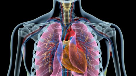
Therefore, stating that the lungs are lateral to the heart means that the lungs are situated to the sides, away from the midline of the body, relative to the heart which is more centrally located in the chest.
The Thoracic Cavity: A Protected Space
The lungs and heart reside within the thoracic cavity, a bony cage formed by the ribs, sternum, and thoracic vertebrae. This cavity provides crucial protection to these vital organs. The thoracic cavity is further subdivided into several compartments, influencing the positioning and function of the heart and lungs.
The Mediastinum: Home to the Heart
The mediastinum is a central compartment within the thoracic cavity, separating the lungs into right and left sides. Importantly, the heart is located within the mediastinum, situated slightly to the left of the midline. This central location allows the heart to effectively pump blood to both the right and left sides of the body.
The Pleural Cavities: Encasing the Lungs
The lungs, on the other hand, occupy the pleural cavities, which are located on either side of the mediastinum. Each lung is surrounded by a double-layered membrane called the pleura. The visceral pleura adheres directly to the lung surface, while the parietal pleura lines the thoracic wall. The space between these two layers is the pleural cavity, filled with a small amount of lubricating fluid. This fluid minimizes friction during respiration. This arrangement ensures that the lungs can expand and contract efficiently during breathing, while maintaining a protective barrier.
The Spatial Relationship: Lungs Lateral, Heart Medial
Given these anatomical structures and their locations, the statement "the lungs are lateral to the heart" becomes clear. The lungs are situated on either side of the mediastinum, away from the midline, whereas the heart resides centrally within the mediastinum, closer to the body's midline. This lateral positioning of the lungs allows for maximal expansion during inhalation, facilitating efficient gas exchange.
Clinical Significance of the Lateral Position of the Lungs
The lateral position of the lungs relative to the heart has significant clinical implications:
1. Auscultation and Percussion: Diagnosing Lung Conditions
Physicians routinely use auscultation (listening with a stethoscope) and percussion (tapping on the chest wall) to assess lung health. The lateral placement of the lungs makes it relatively easy to access these areas for examination. Auscultation can reveal abnormal breath sounds indicative of conditions like pneumonia, bronchitis, or asthma. Percussion can help determine the presence of fluid or air in the pleural space.
2. Chest Radiography and Computed Tomography (CT) Scans: Imaging the Lungs
Imaging techniques like chest X-rays and CT scans rely heavily on the anatomical position of the lungs. Because the lungs are lateral, these imaging modalities can effectively visualize the entire lung field, identifying abnormalities like tumors, infections, or trauma. The mediastinal location of the heart allows for simultaneous evaluation of both cardiac and pulmonary structures within the same image.
3. Cardiopulmonary Procedures: Accessing the Heart and Lungs
Many cardiopulmonary procedures require precise access to the heart and lungs. The lateral location of the lungs influences the approach chosen for procedures such as thoracentesis (removing fluid from the pleural space) or lung biopsies. The central mediastinal location of the heart dictates the approach for cardiac catheterization or coronary artery bypass grafting.
4. Traumatic Injuries: Assessing Lung and Heart Damage
In cases of trauma to the chest, understanding the relationship between the lungs and heart is vital for assessment and treatment. Injuries affecting one structure may indirectly compromise the other. For example, a penetrating injury to the lung might cause pneumothorax (collapsed lung), potentially impacting the heart's function due to changes in intrathoracic pressure. Conversely, blunt force trauma to the chest may damage both the heart and the lungs simultaneously.
5. Respiratory and Cardiovascular Diseases: Interdependence of Systems
Many respiratory and cardiovascular diseases affect both the lungs and the heart. For instance, heart failure can lead to pulmonary edema (fluid accumulation in the lungs), demonstrating the interdependence of the two systems. Conversely, severe lung disease can strain the heart, leading to cor pulmonale (right-sided heart failure). The close proximity of the two organs in the thoracic cavity highlights this intertwined relationship.
Understanding the Complexity: Beyond Simple Laterality
While the simple statement "the lungs are lateral to the heart" provides a fundamental understanding of their positions, the actual relationship is much more nuanced. The heart isn't perfectly centered; it's slightly rotated and tilted within the mediastinum. Similarly, the lungs aren't perfectly symmetrical, with the right lung typically being slightly larger than the left to accommodate the liver. These variations are crucial to remember when interpreting medical images and understanding the clinical presentations of cardiopulmonary conditions.
Conclusion: A Foundational Understanding for Healthcare Professionals
Understanding the spatial relationship between the lungs and the heart—that the lungs are lateral to the heart—is a foundational concept in anatomy and physiology. This knowledge is essential for healthcare professionals, including physicians, nurses, respiratory therapists, and radiologic technologists. This accurate spatial understanding is crucial for proper diagnosis, treatment planning, and overall patient care. The anatomical position of these organs and their relationship to other thoracic structures directly impacts diagnostic procedures, surgical approaches, and the interpretation of medical images. Beyond the basic understanding, appreciating the intricacies and subtle variations in the positioning of the lungs and heart allows for a more comprehensive and accurate assessment of patient health. This article provides a detailed exploration of this vital anatomical relationship and its importance in clinical practice.
Latest Posts
Latest Posts
-
What Would Be The Effect Of A Reduced Venous Return
Apr 06, 2025
-
Iso 9000 Seeks Standardization In Terms Of
Apr 06, 2025
-
If Xy 2 X 2 Y 5 Then Dy Dx
Apr 06, 2025
-
Below Is The Lewis Structure Of The Formaldehyde Molecule
Apr 06, 2025
-
Art Labeling Activity Anatomy And Histology Of The Thyroid Gland
Apr 06, 2025
Related Post
Thank you for visiting our website which covers about The Lungs Are Lateral To The Heart . We hope the information provided has been useful to you. Feel free to contact us if you have any questions or need further assistance. See you next time and don't miss to bookmark.
