The Contractile Molecules In Muscle Cells Are Blank______.
Holbox
Mar 31, 2025 · 6 min read

Table of Contents
- The Contractile Molecules In Muscle Cells Are Blank______.
- Table of Contents
- The Contractile Molecules in Muscle Cells are Actin and Myosin
- Understanding the Muscle Cell Structure: A Microscopic View
- The Sarcomere: The Engine of Muscle Contraction
- Actin: The Thin Filament's Backbone
- Myosin: The Molecular Motor
- The Sliding Filament Theory: Unifying Actin and Myosin's Action
- Muscle Contraction: A Symphony of Molecules
- Different Muscle Types: Variations on a Theme
- Skeletal Muscle: Voluntary Movement
- Cardiac Muscle: The Heart's Rhythm
- Smooth Muscle: Involuntary Control
- Muscle Relaxation: Reversing the Process
- Diseases and Disorders Affecting Muscle Contraction
- Conclusion: Actin and Myosin: The Cornerstones of Movement
- Latest Posts
- Latest Posts
- Related Post
The Contractile Molecules in Muscle Cells are Actin and Myosin
Muscle contraction, that fundamental process enabling movement, from a heartbeat to a marathon run, hinges on the intricate interplay of specific proteins within muscle cells. The answer to the question, "The contractile molecules in muscle cells are blank______," is unequivocally actin and myosin. These two proteins, along with a supporting cast of regulatory and structural molecules, orchestrate the remarkable mechanics of muscle function. This article delves deep into the structure and function of actin and myosin, exploring their roles in various muscle types and the mechanisms driving muscle contraction and relaxation.
Understanding the Muscle Cell Structure: A Microscopic View
Before exploring the molecular players, it's crucial to understand the cellular context. Muscle cells, or myocytes, are highly specialized cells packed with long, cylindrical structures called myofibrils. These myofibrils are the functional units of muscle contraction, appearing under a microscope as repeating units called sarcomeres. The sarcomere is the fundamental structural and functional unit of striated muscle tissue (skeletal and cardiac muscle), responsible for the characteristic striated appearance due to the organized arrangement of actin and myosin filaments.
The Sarcomere: The Engine of Muscle Contraction
The sarcomere is delimited by Z-lines, which serve as anchoring points for the thin filaments (primarily composed of actin). Between the Z-lines, thick filaments (primarily composed of myosin) interdigitate with the thin filaments. This precise arrangement allows for the sliding filament mechanism, the core process driving muscle contraction.
Actin: The Thin Filament's Backbone
Actin, a globular protein (G-actin), polymerizes to form long, fibrous strands called F-actin. These F-actin strands are intertwined to create the thin filaments. Several other proteins are associated with the thin filaments, playing vital roles in regulating muscle contraction. These include:
- Tropomyosin: A long, fibrous protein that winds around the F-actin strand, covering the myosin-binding sites on actin in a relaxed muscle.
- Troponin: A complex of three proteins (troponin I, T, and C) that binds to tropomyosin and actin. Troponin C binds calcium ions, triggering a conformational change that shifts tropomyosin, exposing the myosin-binding sites on actin.
This intricate interplay of actin, tropomyosin, and troponin is crucial for regulating muscle contraction and ensuring it occurs only when necessary. The presence of calcium ions is essential for initiating the process.
Myosin: The Molecular Motor
Myosin is a motor protein responsible for generating the force necessary for muscle contraction. It's a large, complex protein consisting of two heavy chains and several light chains. The heavy chains form a long, rod-like tail and two globular heads. The myosin heads possess ATPase activity, meaning they can hydrolyze ATP (adenosine triphosphate), releasing energy that drives the interaction with actin.
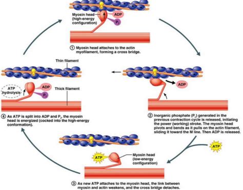
The myosin heads bind to actin, forming cross-bridges. The cyclical process of cross-bridge cycling involves:
- ATP Binding: Myosin heads bind to ATP, causing them to detach from actin.
- ATP Hydrolysis: ATP is hydrolyzed to ADP and inorganic phosphate (Pi), causing a conformational change in the myosin head, cocking it into a high-energy state.
- Cross-Bridge Formation: The cocked myosin head binds to a new site on the actin filament.
- Power Stroke: The release of ADP and Pi triggers the power stroke, a conformational change in the myosin head that pulls the actin filament towards the center of the sarcomere.
- ADP Release: ADP is released, and the myosin head remains bound to actin until another ATP molecule binds.
This cycle repeats numerous times during a single contraction, resulting in the sliding of actin and myosin filaments past each other, shortening the sarcomere and ultimately causing muscle contraction.
The Sliding Filament Theory: Unifying Actin and Myosin's Action
The sliding filament theory elegantly explains how actin and myosin interaction leads to muscle contraction. The theory posits that muscle contraction occurs due to the relative sliding of actin and myosin filaments past each other, without the filaments themselves shortening. The myosin heads, fueled by ATP hydrolysis, repeatedly bind to actin, perform power strokes, and detach, pulling the actin filaments towards the center of the sarcomere. This process continues until the muscle is fully contracted.
Muscle Contraction: A Symphony of Molecules
Muscle contraction isn't solely dependent on actin and myosin; other proteins play crucial roles. Titin, a giant protein, acts as a molecular spring, providing elasticity and stability to the sarcomere. Nebulin, another structural protein, helps align the thin filaments. Furthermore, the sarcoplasmic reticulum, a specialized endoplasmic reticulum within muscle cells, stores and releases calcium ions, a critical trigger for initiating muscle contraction.
Different Muscle Types: Variations on a Theme
While actin and myosin are fundamental to all muscle types, variations exist in their organization and regulatory mechanisms.
Skeletal Muscle: Voluntary Movement
Skeletal muscle, responsible for voluntary movement, is characterized by its highly organized, striated appearance due to the regular arrangement of sarcomeres. Contraction is rapid and powerful, driven by the precise coordination of actin, myosin, and the associated regulatory proteins.
Cardiac Muscle: The Heart's Rhythm
Cardiac muscle, found in the heart, exhibits striations similar to skeletal muscle but possesses unique features. It's capable of spontaneous contraction, regulated by specialized pacemaker cells. Intercalated discs, connecting adjacent cardiac muscle cells, enable rapid and synchronized contraction. Actin and myosin are essential for contraction, but the regulatory mechanisms differ slightly from skeletal muscle.
Smooth Muscle: Involuntary Control
Smooth muscle, found in the walls of internal organs and blood vessels, lacks the striated appearance of skeletal and cardiac muscle. Its contraction is slower and more sustained, controlled by different signaling pathways and regulatory proteins compared to striated muscles. Actin and myosin are still the primary contractile proteins, but their organization and regulation are less structured.
Muscle Relaxation: Reversing the Process
Muscle relaxation involves the reversal of the contraction process. Calcium ions are actively pumped back into the sarcoplasmic reticulum by calcium ATPases, reducing calcium concentration in the cytosol. This decreased calcium concentration allows tropomyosin to return to its resting position, blocking the myosin-binding sites on actin, thereby preventing further cross-bridge formation and halting muscle contraction.
Diseases and Disorders Affecting Muscle Contraction
Dysfunctions in actin, myosin, or other associated proteins can lead to various muscle diseases and disorders. These include muscular dystrophies, characterized by progressive muscle weakness and degeneration; myasthenia gravis, an autoimmune disorder affecting neuromuscular junctions; and various cardiomyopathies affecting heart muscle function. Understanding the molecular mechanisms of muscle contraction is crucial for developing effective treatments for these conditions.
Conclusion: Actin and Myosin: The Cornerstones of Movement
In conclusion, the contractile molecules in muscle cells are definitively actin and myosin. Their intricate interaction, governed by a complex interplay of regulatory proteins and calcium ions, underlies the remarkable ability of muscles to contract and relax, enabling movement, maintaining posture, and performing a myriad of vital functions throughout the body. Further research into the intricacies of this process promises to yield advancements in understanding and treating muscle-related diseases and enhancing human performance. The exploration of actin and myosin extends far beyond simple textbook descriptions; it’s a journey into the very essence of movement and life itself. Understanding their roles, their interplay, and the cellular environment that supports them provides an unparalleled insight into the elegant mechanics of the human body.
Latest Posts
Latest Posts
-
Which Code Example Is An Expression
Apr 03, 2025
-
Good Team Goals Are All Of The Following Except
Apr 03, 2025
-
During The Year Trc Corporation Has The Following Inventory Transactions
Apr 03, 2025
-
In Project Network Analysis Slack Refers To The Difference Between
Apr 03, 2025
-
The Economic Way Of Thinking Stresses That
Apr 03, 2025
Related Post
Thank you for visiting our website which covers about The Contractile Molecules In Muscle Cells Are Blank______. . We hope the information provided has been useful to you. Feel free to contact us if you have any questions or need further assistance. See you next time and don't miss to bookmark.
