Procedure 4 Testing The Extrinsic Eye Muscles
Holbox
Apr 01, 2025 · 6 min read
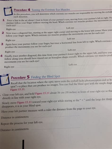
Table of Contents
- Procedure 4 Testing The Extrinsic Eye Muscles
- Table of Contents
- Procedures for Testing Extrinsic Eye Muscles: A Comprehensive Guide
- Understanding the Extrinsic Eye Muscles
- Methods for Testing Extrinsic Eye Muscles
- 1. Assessing Visual Acuity
- 2. Cover Test
- 3. Cardinal Positions of Gaze
- 4. Ductions and Versions
- 5. Measurement of Eye Movements
- Interpreting the Results
- Differential Diagnosis
- Conclusion
- Latest Posts
- Latest Posts
- Related Post
Procedures for Testing Extrinsic Eye Muscles: A Comprehensive Guide
The extrinsic eye muscles are six muscles responsible for the movement and positioning of the eyes. Accurate assessment of their function is crucial for diagnosing a wide range of ophthalmological and neurological conditions. This article provides a comprehensive guide to the procedures used for testing these vital muscles, detailing the methods, expected findings, and interpretation of results. Understanding these procedures is essential for healthcare professionals involved in ophthalmology, neurology, and optometry.
Understanding the Extrinsic Eye Muscles
Before delving into testing procedures, it's vital to understand the six extrinsic eye muscles and their respective actions:
- Medial Rectus: Adducts (turns the eye inward) the eye.
- Lateral Rectus: Abducts (turns the eye outward) the eye.
- Superior Rectus: Elevates (turns the eye upward), intorts (rotates the top of the eye towards the nose), and adducts the eye.
- Inferior Rectus: Depresses (turns the eye downward), extorts (rotates the top of the eye away from the nose), and adducts the eye.
- Superior Oblique: Intorts, depresses, and abducts the eye.
- Inferior Oblique: Extorts, elevates, and abducts the eye.
Understanding the coordinated action of these muscles is key to interpreting the results of any testing. Dysfunction in one or more of these muscles can lead to strabismus (misalignment of the eyes), diplopia (double vision), or other visual disturbances.
Methods for Testing Extrinsic Eye Muscles
Several procedures are used to evaluate the function of the extrinsic eye muscles. These methods assess both the strength and coordination of these muscles.
1. Assessing Visual Acuity
Before initiating any muscle testing, it's crucial to establish the patient's visual acuity. This baseline measurement helps to differentiate between muscle weakness and visual impairment contributing to any observed eye movement abnormalities. Standard visual acuity charts (Snellen chart or similar) are used for this purpose. Correcting lenses should be used if the patient is habitually spectacle-wearing.
2. Cover Test
The cover test is a fundamental test for detecting strabismus (eye misalignment) and assessing the presence of phorias (latent deviations of the eyes that are overcome by fusion). The procedure involves:
- Alternate Cover Test: The examiner alternately covers one eye at a time while observing the uncovered eye for any movement. A movement of the uncovered eye to re-fixate on the target indicates a phoria or tropia (manifest strabismus).
- Simultaneous Cover Test: Both eyes are covered briefly and then uncovered simultaneously. This test helps assess fusion and suppression (the brain's suppression of the image from one eye to avoid diplopia).
Interpretation: Movement of the uncovered eye indicates a deviation. The direction of the movement helps identify the affected muscle(s). For example, a movement outward (abduction) suggests a possible medial rectus weakness, and inward (adduction) movement suggests a possible lateral rectus weakness.
3. Cardinal Positions of Gaze
This method evaluates the action of each muscle in isolation by moving the patient's gaze through nine cardinal positions of gaze. The patient is instructed to follow the examiner's finger or a light source as it moves through the following positions:
- Primary Position: Looking straight ahead.
- Right Gaze: Looking to the right.
- Left Gaze: Looking to the left.
- Upward Gaze: Looking upwards.
- Downward Gaze: Looking downwards.
- Up and Right Gaze: Looking up and to the right.
- Up and Left Gaze: Looking up and to the left.
- Down and Right Gaze: Looking down and to the right.
- Down and Left Gaze: Looking down and to the left.
Observation: The examiner observes the eyes for any limitations in movement, nystagmus (involuntary eye movements), or any other abnormal movements in each position. This helps to identify the muscle responsible for the movement in the particular gaze position. Weakness or paralysis of a muscle will be evident as a limitation in movement towards the relevant direction.
4. Ductions and Versions
This test differentiates between the actions of individual muscles (ductions) and the synergistic action of muscles in both eyes (versions):
- Ductions: The examiner assesses each eye's movement independently, covering the other eye to isolate the action. This helps to observe individual muscle strength and function.
- Versions: Both eyes are observed simultaneously as the patient follows the examiner's finger or light source through the nine cardinal positions of gaze. This assesses the coordination between the eyes and the synergistic action of different muscles.
Interpretation: Limitations or dyscoordination during ductions or versions point towards a specific muscle weakness or neurological impairment.
5. Measurement of Eye Movements
Precise measurements of eye movements can be obtained using sophisticated instruments like:
- Electro-oculography (EOG): This technique measures the corneal-retinal potential to track eye movements.
- Videooculography (VOG): This involves recording eye movements using a video camera.
- Infrared Oculography: This uses infrared light to track pupil movements with high precision.
These instruments provide objective data on the speed, amplitude, and accuracy of eye movements, providing a more quantitative assessment compared to visual observation. They are particularly useful in evaluating subtle motility disorders or in research settings.
Interpreting the Results
The interpretation of the results from these tests requires a thorough understanding of the anatomy and physiology of the extrinsic eye muscles. The following are some key considerations:
- Limitation of gaze: A restriction in gaze in a specific direction suggests weakness or paralysis of the muscle primarily responsible for movement in that direction.
- Diplopia: Double vision, often occurring in specific gaze positions, indicates a disconjugate gaze (misalignment of the eyes).
- Nystagmus: Involuntary rhythmic oscillations of the eyes suggest a neurological problem affecting the oculomotor pathways.
- Ptosis (drooping eyelid): This can indicate a problem with the oculomotor nerve (CN III).
- Asymmetry of muscle action: Differences in the strength or range of motion between the two eyes suggest muscle imbalance.
Specific Muscle Weakness Manifestations:
- Medial Rectus Weakness: Difficulty adducting the eye, causing exotropia (outward turning of the eye).
- Lateral Rectus Weakness: Difficulty abducting the eye, causing esotropia (inward turning of the eye).
- Superior Rectus Weakness: Difficulty elevating and adducting the eye.
- Inferior Rectus Weakness: Difficulty depressing and adducting the eye.
- Superior Oblique Weakness: Difficulty intorting, depressing, and abducting the eye, often resulting in hypertropia (one eye positioned higher than the other).
- Inferior Oblique Weakness: Difficulty extorting, elevating, and abducting the eye, often leading to hypotropia (one eye positioned lower than the other).
Differential Diagnosis
Abnormal results from extrinsic eye muscle testing require a thorough differential diagnosis to identify the underlying cause. Possible causes include:
- Myopathies: Muscle diseases affecting the extrinsic eye muscles.
- Neuropathies: Conditions affecting the cranial nerves that innervate the eye muscles (CN III, IV, VI).
- Trauma: Injury to the eye or surrounding structures.
- Congenital abnormalities: Birth defects affecting the development of eye muscles.
- Neurological disorders: Conditions like stroke, multiple sclerosis, or myasthenia gravis can affect eye movements.
- Orbital diseases: Tumors or other conditions within the orbit can affect eye muscle function.
A detailed history, comprehensive neurological examination, and often additional investigations such as imaging studies (MRI or CT scans) are essential for accurate diagnosis.
Conclusion
Testing the extrinsic eye muscles is a crucial component of ophthalmological and neurological examinations. The combination of clinical tests, such as the cover test and assessment of cardinal positions of gaze, along with advanced techniques such as EOG and VOG, provides a comprehensive evaluation of eye movement function. Accurate interpretation of these tests, combined with a thorough differential diagnosis, is essential for the timely detection and appropriate management of various ophthalmological and neurological conditions impacting eye motility. This detailed understanding of the procedures and their implications empowers healthcare professionals to provide optimal patient care.
Latest Posts
Latest Posts
-
Rank The Indicated Protons In Decreasing Order Of Acidity
Apr 05, 2025
-
The Drawing Shows A Hydraulic Chamber With A Spring
Apr 05, 2025
-
A Market Constraint Can Be Overcome By
Apr 05, 2025
-
Highest Temp For Cold Holding Tuna Salad
Apr 05, 2025
-
Biochemistry A Short Course 4th Edition
Apr 05, 2025
Related Post
Thank you for visiting our website which covers about Procedure 4 Testing The Extrinsic Eye Muscles . We hope the information provided has been useful to you. Feel free to contact us if you have any questions or need further assistance. See you next time and don't miss to bookmark.
