Light Source Of A Microscope Function
Holbox
Mar 10, 2025 · 7 min read
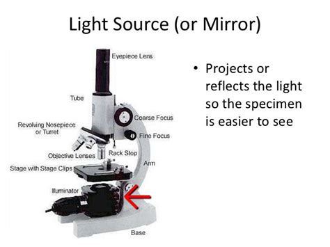
Table of Contents
Understanding the Light Source of a Microscope: Function, Types, and Optimization
The microscope, a cornerstone of scientific discovery, wouldn't be possible without its crucial component: the light source. The function of the light source extends far beyond simply illuminating the specimen; it dictates the quality of the image, the techniques that can be employed, and ultimately, the conclusions drawn from microscopic observation. This in-depth exploration delves into the intricacies of microscope light sources, covering their function, various types, and optimization strategies for achieving optimal imaging results.
The Fundamental Role of Illumination in Microscopy
Before delving into specific light sources, it's crucial to understand the overarching function of illumination in microscopy. The light source's primary role is to provide sufficient and appropriately controlled illumination to allow visualization of the specimen. However, this seemingly simple function encompasses several critical aspects:
1. Specimen Illumination: Achieving Sufficient Brightness and Evenness
The light source must provide sufficient brightness to illuminate the specimen adequately. Insufficient light results in a dim, hard-to-interpret image, while excessive light can lead to bleaching of fluorescent samples or damage to delicate specimens. Furthermore, the illumination needs to be even across the entire field of view. Uneven illumination leads to variations in brightness across the image, making it difficult to assess details accurately. This is particularly important in quantitative microscopy where precise measurements are required.
2. Contrast Enhancement: Revealing Fine Details
The human eye struggles to perceive fine details in specimens lacking sufficient contrast. The light source plays a critical role in enhancing contrast. Different microscopy techniques, such as brightfield, darkfield, phase contrast, and fluorescence, rely on specific manipulation of light to enhance contrast in unique ways. The light source's characteristics—wavelength, intensity, and coherence—directly impact the success of these contrast-enhancing techniques.
3. Excitation of Fluorophores (Fluorescence Microscopy): Enabling Specific Visualization
In fluorescence microscopy, the light source doesn't merely illuminate the specimen; it excites fluorophores (fluorescent molecules) within the specimen. These fluorophores then emit light at a longer wavelength, allowing visualization of specific structures or molecules labeled with these fluorescent probes. The choice of light source is crucial here as the excitation wavelength must match the absorption spectrum of the specific fluorophore.
4. Minimizing Phototoxicity and Bleaching: Protecting the Specimen
Prolonged exposure to intense light can damage the specimen through phototoxicity (damage caused by light) or bleaching (loss of fluorescence). The light source's intensity and exposure time must be carefully controlled to minimize these effects, particularly in live-cell imaging. This often requires the use of specialized filters, low-intensity light sources, and rapid image acquisition techniques.
Types of Microscope Light Sources
Microscope light sources have evolved significantly over time, with various types now available, each with its advantages and disadvantages.
1. Tungsten-Halogen Lamps: The Traditional Workhorse
Tungsten-Halogen lamps are incandescent lamps that produce a continuous spectrum of visible light. They are relatively inexpensive, easy to use, and readily available. However, they generate significant heat, have a shorter lifespan compared to other modern light sources, and emit less intense light in the blue and UV regions of the spectrum. They're still commonly used in basic brightfield microscopy but are gradually being replaced by more efficient options.
2. LED (Light-Emitting Diode) Light Sources: The Modern Standard
LEDs have become the dominant light source in modern microscopy. They offer several advantages:
- Long lifespan: LEDs have significantly longer lifespans than tungsten-halogen lamps, reducing the need for frequent replacements.
- Energy efficiency: LEDs are much more energy-efficient, leading to lower operational costs and reduced heat generation.
- Compact size: Their small size makes them ideal for integration into various microscope designs.
- Color control: LEDs are available in a wide range of colors and can be easily adjusted for intensity. This allows for precise control over excitation wavelength in fluorescence microscopy.
- Rapid switching: LEDs can be switched on and off very quickly, making them suitable for time-lapse imaging and other dynamic applications.
3. Laser Light Sources: High Intensity and Coherence for Specialized Applications
Lasers provide highly intense, monochromatic (single wavelength) light. Their high intensity is crucial for applications like confocal microscopy and super-resolution microscopy, where high signal-to-noise ratios are essential. However, lasers are more expensive than LEDs and require careful handling due to potential eye damage. Laser light's coherence (all light waves in phase) can lead to speckle artifacts in some imaging techniques, requiring mitigation strategies.
4. Xenon Arc Lamps: Powerful Illumination for Fluorescence Microscopy
Xenon arc lamps are high-intensity light sources that produce a continuous spectrum covering both visible and ultraviolet light. This makes them suitable for fluorescence microscopy, particularly for applications requiring excitation across a broad range of wavelengths. However, they are less energy-efficient than LEDs, have a shorter lifespan, and generate significant heat. They are increasingly being replaced by high-power LED arrays for fluorescence applications.
5. Mercury Arc Lamps: A Legacy Source for Fluorescence Microscopy
Mercury arc lamps, while largely superseded by Xenon and high-power LEDs, are older high-intensity light sources used in fluorescence microscopy. They produce specific emission lines that can be used for excitation of various fluorophores. However, they have significant disadvantages including shorter lifespan, high heat output, and the need for specialized power supplies.
Optimizing Microscope Light Source Performance
Achieving optimal results with any microscope light source requires careful consideration of several factors:
1. Light Intensity Control: Finding the "Sweet Spot"
The light intensity should be adjusted to provide sufficient illumination for the specimen without causing damage or bleaching. This often involves a delicate balance, requiring careful observation and experimentation. Many modern microscopes allow for precise control of light intensity, enabling optimization for each sample and imaging technique.
2. Kohler Illumination: Ensuring Even and Optimal Illumination
Kohler illumination is a crucial technique for achieving even and optimal illumination across the field of view. It involves adjusting the condenser and field diaphragm to correctly focus the light source onto the specimen. This minimizes artifacts and maximizes image quality. Mastering Kohler illumination is essential for achieving consistent and high-quality images.
3. Filter Selection: Fine-tuning the Wavelength and Intensity
Filters play a vital role in shaping the light reaching the specimen. In fluorescence microscopy, excitation filters select the appropriate wavelength for exciting fluorophores, while emission filters block out unwanted light and isolate the emitted fluorescence. In other techniques, filters can be used to enhance contrast or adjust the overall color balance.
4. Light Path Alignment: Ensuring Proper Light Delivery
A properly aligned light path is critical for optimal performance. This involves ensuring that the light source is correctly focused and centered on the condenser and that the condenser is properly adjusted to provide appropriate illumination for the chosen objective. Misalignment can lead to uneven illumination, reduced resolution, and artifacts.
5. Regular Maintenance: Extending Lifespan and Performance
Regular maintenance, such as cleaning the light source and filters, is essential for maintaining optimal performance and extending the lifespan of the components. Dust and other contaminants can significantly reduce light intensity and quality. Following the manufacturer's recommendations for maintenance is critical.
Conclusion: The Light Source – A Cornerstone of Microscopy's Success
The light source isn't merely an accessory in a microscope; it's an integral component that fundamentally dictates image quality and the applicability of various microscopy techniques. Understanding its function, the diverse types available, and how to optimize its performance is vital for achieving optimal results in microscopy. From the traditional tungsten-halogen lamps to the modern prevalence of LEDs and the specialized applications of lasers, the ongoing evolution of microscope light sources continually pushes the boundaries of what's possible in microscopic observation and scientific discovery. The selection and optimization of the light source are critical steps in ensuring high-quality, reliable, and informative microscopic imaging.
Latest Posts
Latest Posts
-
The Average Variable Cost Per Sale At H
Mar 10, 2025
-
Low Is Too High As Easy Is To
Mar 10, 2025
-
If Is A Linear Transformation Such That And Then
Mar 10, 2025
-
Molar Latent Heat Of The Transformation Caf2
Mar 10, 2025
-
When Approaching A Merging Traffic Sign You Should
Mar 10, 2025
Related Post
Thank you for visiting our website which covers about Light Source Of A Microscope Function . We hope the information provided has been useful to you. Feel free to contact us if you have any questions or need further assistance. See you next time and don't miss to bookmark.
