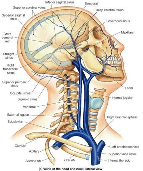
Label The Veins Of The Head And Neck
Holbox
Mar 31, 2025 · 6 min read

Table of Contents
- Label The Veins Of The Head And Neck
- Table of Contents
- Labeling the Veins of the Head and Neck: A Comprehensive Guide
- Major Veins of the Head and Neck: A Systematic Approach
- Superficial Veins
- Deep Veins
- Clinical Correlations and Significance
- Mnemonic Devices for Remembering the Veins
- Conclusion
- Latest Posts
- Latest Posts
- Related Post
Labeling the Veins of the Head and Neck: A Comprehensive Guide
The venous system of the head and neck is a complex network responsible for draining deoxygenated blood from the brain, face, and neck back to the heart. Understanding its intricate anatomy is crucial for medical professionals, students, and anyone interested in human physiology. This comprehensive guide will explore the major veins of this region, detailing their locations, tributaries, and clinical significance. We'll delve into the intricacies of this vascular network, providing a detailed roadmap for navigating the veins of the head and neck.
Major Veins of the Head and Neck: A Systematic Approach
The venous drainage of the head and neck can be broadly categorized into superficial and deep systems. These systems, while distinct, are interconnected, ensuring efficient blood return to the heart.
Superficial Veins
The superficial veins are located closer to the skin surface and are often visible. They play a vital role in draining blood from the scalp, face, and superficial tissues of the neck.
1. External Jugular Vein: This prominent vein is easily palpable along the sternocleidomastoid muscle in the neck. It arises from the posterior facial vein and receives tributaries from the occipital region and posterior auricular vein before emptying into the subclavian vein. Its superficial location makes it a readily accessible site for venipuncture.
Clinical Significance: Distension of the external jugular vein can indicate increased central venous pressure, a sign of potential heart failure or other cardiovascular issues.
2. Internal Jugular Vein: A much larger and deeper vein than its external counterpart, the internal jugular vein drains blood from the brain, face, and neck. It descends alongside the carotid artery in the carotid sheath. It's formed by the confluence of the sigmoid sinus (from the brain) and receives numerous tributaries.
Clinical Significance: Obstruction of the internal jugular vein can lead to significant cerebral edema and other neurological complications. It's also a critical site for central venous catheterization.
3. Facial Vein: This vein drains blood from the face and communicates with the cavernous sinus, a crucial intracranial venous structure. It is formed by the union of the angular and supraorbital veins.
Clinical Significance: Infections originating in the face, especially in the danger triangle of the face (the area between the eyebrows and upper lip), can spread via the facial vein to the cavernous sinus, leading to a potentially life-threatening condition known as cavernous sinus thrombosis.
4. Anterior Jugular Vein: This smaller vein runs along the midline of the neck, draining blood from the anterior neck region. It often communicates with its contralateral counterpart and the external jugular vein before emptying into the subclavian vein.
Clinical Significance: While not as clinically significant as the internal or external jugular veins, its role in draining the anterior neck region is important.
5. Posterior Auricular Vein: Located behind the ear, this vein drains blood from the scalp and posterior auricular region. It joins the external jugular vein.
6. Occipital Vein: Drains blood from the occipital region of the scalp and usually joins the external jugular vein.
7. Superficial Temporal Vein: Drains the superficial temporal region of the scalp and joins the maxillary vein to form the retromandibular vein.
8. Maxillary Vein: Formed by the pterygoid plexus and drains the deep structures of the face and joins the superficial temporal vein to form the retromandibular vein.
9. Retromandibular Vein: This vein is formed by the confluence of the superficial temporal and maxillary veins. It then divides into anterior and posterior divisions. The anterior division joins the facial vein, while the posterior division joins the external jugular vein.
Deep Veins
The deep veins of the head and neck are located deeper within the tissues and are often associated with arteries and nerves within specific fascial compartments.
1. Vertebral Vein: This vein drains blood from the vertebral column and its associated muscles. It runs alongside the vertebral artery and ultimately empties into the brachiocephalic vein.
Clinical Significance: Vertebral vein thrombosis is a rare but serious condition that can cause significant neurological deficits.
2. Internal Vertebral Plexus: A network of veins surrounding the vertebral column, contributing to the vertebral vein.
3. Pharyngeal Veins: These veins drain the pharynx and often communicate with other veins in the neck.
4. Pterygoid Plexus: A complex network of veins located in the infratemporal fossa, draining the muscles of mastication. It contributes to the maxillary vein.
5. Cavernous Sinus: This is a crucial intracranial venous structure located within the sella turcica. It receives blood from the ophthalmic veins and drains into the internal jugular vein.
Clinical Significance: Due to its proximity to the brain and its connections to other venous structures, the cavernous sinus is susceptible to infections, leading to conditions such as cavernous sinus thrombosis.
Clinical Correlations and Significance
Understanding the venous anatomy of the head and neck is essential in various medical contexts:
-
Venipuncture: The external jugular vein is often used for venipuncture due to its superficial location and large size.
-
Central Venous Catheterization: The internal jugular vein is a common site for inserting central venous catheters, allowing for intravenous access to the central circulation.
-
Stroke and Cerebral Edema: Obstruction or impaired drainage of the venous system can significantly contribute to stroke and cerebral edema.
-
Infections: Infections in the face and neck can spread through the venous system, leading to life-threatening complications like cavernous sinus thrombosis.
-
Surgical Procedures: Knowledge of the venous anatomy is crucial for surgeons performing procedures in the head and neck region to avoid damaging these vital vessels.
-
Imaging Interpretation: Radiological images such as venograms and CT scans often require a thorough understanding of the venous anatomy for accurate interpretation.
Mnemonic Devices for Remembering the Veins
Remembering the intricate network of veins can be challenging. Using mnemonic devices can significantly improve recall. Here are a few examples:
-
For the superficial veins: Think of the acronym E-F-A-I-J (External Jugular, Facial, Anterior Jugular, Internal Jugular).
-
For the deep veins: Focus on their location – V-P-C (Vertebral, Pterygoid Plexus, Cavernous Sinus).
While these are simplified mnemonics, they serve as starting points for memorization. Active recall and repeated study are crucial for solidifying your understanding.
Conclusion
The venous system of the head and neck is a complex and vital part of the circulatory system. Understanding its anatomy and clinical significance is crucial for healthcare professionals, medical students, and anyone with an interest in human physiology. By studying the major veins, their tributaries, and their clinical implications, we gain a deeper appreciation for the intricate workings of this essential network. This guide provides a comprehensive overview, laying the groundwork for a more profound understanding of this complex vascular territory. Remember that continued study and exploration are key to mastering the intricacies of this anatomical region. Consult anatomical atlases and relevant textbooks for further detailed information and visualization.
Latest Posts
Latest Posts
-
Water Flows Steadily From A Tank Mounted On A Cart
Apr 03, 2025
-
Match The Accounting Terms With The Corresponding Definitions
Apr 03, 2025
-
The Diagram Shows The Reactions Of The Beta Oxidation Pathway
Apr 03, 2025
-
Educational Appeals Make The Assumption That
Apr 03, 2025
-
Unlike Firms That Outsource Firms Engaged In Offshoring
Apr 03, 2025
Related Post
Thank you for visiting our website which covers about Label The Veins Of The Head And Neck . We hope the information provided has been useful to you. Feel free to contact us if you have any questions or need further assistance. See you next time and don't miss to bookmark.
