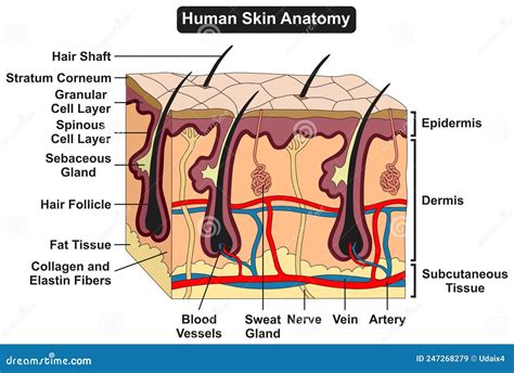
Label The Parts Of The Skin And Subcutaneous Tissue
Holbox
Mar 31, 2025 · 6 min read

Table of Contents
- Label The Parts Of The Skin And Subcutaneous Tissue
- Table of Contents
- Label the Parts of the Skin and Subcutaneous Tissue: A Comprehensive Guide
- The Epidermis: The Outermost Shield
- 1. Stratum Corneum:
- 2. Stratum Lucidum:
- 3. Stratum Granulosum:
- 4. Stratum Spinosum:
- 5. Stratum Basale (Germinativum):
- The Dermis: Strength, Support, and Sensation
- 1. Papillary Layer:
- 2. Reticular Layer:
- The Subcutaneous Tissue (Hypodermis): Insulation and Energy Storage
- 1. Adipose Tissue:
- 2. Connective Tissue:
- Specialized Structures Within the Skin
- 1. Hair Follicles:
- 2. Sebaceous Glands:
- 3. Sweat Glands (Sudoriferous Glands):
- 4. Sensory Receptors:
- 5. Blood Vessels:
- 6. Lymph Vessels:
- 7. Nails:
- Clinical Significance
- Conclusion
- Latest Posts
- Latest Posts
- Related Post
Label the Parts of the Skin and Subcutaneous Tissue: A Comprehensive Guide
The skin, our largest organ, is a remarkable structure, a complex and dynamic barrier protecting us from the external environment. Understanding its intricate layers and the underlying subcutaneous tissue is crucial for appreciating its diverse functions and the impact of various dermatological conditions. This comprehensive guide will delve into the detailed anatomy of the skin and subcutaneous tissue, labeling its key components and explaining their roles.
The Epidermis: The Outermost Shield
The epidermis, the outermost layer of the skin, is a stratified squamous epithelium, meaning it's composed of multiple layers of flattened cells. Its primary function is protection. Let's break down its key components:
1. Stratum Corneum:
- Function: The outermost layer, composed of dead, keratinized cells (corneocytes). This acts as a tough, waterproof barrier, protecting against dehydration, abrasion, and pathogen entry. Its constant shedding (desquamation) ensures continuous renewal.
2. Stratum Lucidum:
- Function: A thin, translucent layer found only in thick skin (palms and soles). Composed of flattened, densely packed keratinocytes, it contributes to the skin's barrier function and light refraction.
3. Stratum Granulosum:
- Function: Cells in this layer begin to die as they lose their nuclei and organelles. Keratohyalin granules appear, contributing to keratinization, the process of hardening the cells. This layer plays a vital role in water retention and barrier formation.
4. Stratum Spinosum:
- Function: Characterized by spiny-looking cells connected by desmosomes, strong intercellular junctions. This layer contains Langerhans cells, crucial for immune responses against pathogens. Cell division contributes to epidermal renewal.
5. Stratum Basale (Germinativum):
- Function: The deepest layer, resting on the basement membrane separating the epidermis from the dermis. This is the site of active cell division (mitosis), generating new keratinocytes that migrate upwards through the other epidermal layers. Melanocytes, responsible for melanin production (pigmentation), are also found here. Merkel cells, involved in touch sensation, are present in this layer.
The Dermis: Strength, Support, and Sensation
The dermis, a much thicker layer than the epidermis, lies beneath it and provides structural support. It's composed primarily of connective tissue, giving the skin its strength and elasticity. Let's examine its key features:
1. Papillary Layer:
- Function: This superficial layer consists of loose connective tissue, forming dermal papillae (finger-like projections) that interdigitate with the epidermis. These papillae increase the surface area for nutrient exchange and enhance the adhesion between the epidermis and dermis. Meissner's corpuscles, responsible for light touch sensation, are located here. Capillary networks supply oxygen and nutrients to the epidermis.
2. Reticular Layer:
- Function: This deeper layer is composed of dense irregular connective tissue, providing the skin's strength and elasticity. Collagen fibers provide tensile strength, while elastin fibers allow for stretching and recoil. This layer contains Pacinian corpuscles, which detect pressure and vibration. Hair follicles, sebaceous glands (oil glands), and sweat glands are embedded within the reticular layer.
The Subcutaneous Tissue (Hypodermis): Insulation and Energy Storage
The subcutaneous tissue, also known as the hypodermis, lies beneath the dermis. It's not technically part of the skin but plays a crucial role in its overall function:
1. Adipose Tissue:
- Function: This is the primary component of the subcutaneous tissue, consisting of fat cells (adipocytes). Adipose tissue acts as an energy reservoir, insulation against cold, and cushioning against impact. Its thickness varies significantly depending on body location and individual factors.
2. Connective Tissue:
- Function: Connective tissue, including collagen and elastin fibers, provides structural support and connects the skin to underlying muscles and bones. Blood vessels and nerves traverse this layer, supplying the skin and subcutaneous tissue.
Specialized Structures Within the Skin
Several specialized structures are integral to the skin's function:
1. Hair Follicles:
- Function: These are tubular invaginations of the epidermis extending deep into the dermis. They produce hair, providing insulation and protection. Each follicle contains a hair bulb, where hair growth originates, and arrector pili muscles, which cause hair to stand on end (goosebumps).
2. Sebaceous Glands:
- Function: These are associated with hair follicles and secrete sebum, an oily substance that lubricates the skin and hair, preventing dryness and providing a barrier against pathogens.
3. Sweat Glands (Sudoriferous Glands):
- Function: These glands produce sweat, contributing to thermoregulation (body temperature control) and excretion of waste products. Eccrine sweat glands are widely distributed and secrete a watery fluid, while apocrine sweat glands are located in specific areas (armpits, groin) and secrete a thicker, odorous fluid.
4. Sensory Receptors:
- Function: Various sensory receptors, including Meissner's corpuscles (light touch), Pacinian corpuscles (pressure and vibration), and free nerve endings (pain, temperature), are embedded within the dermis and subcutaneous tissue, providing the body with sensory information about its surroundings.
5. Blood Vessels:
- Function: A dense network of blood vessels within the dermis and subcutaneous tissue supplies the skin with oxygen and nutrients and removes waste products. They also play a crucial role in thermoregulation by controlling blood flow to the skin's surface.
6. Lymph Vessels:
- Function: A network of lymph vessels within the dermis and subcutaneous tissue aids in immune system function by collecting and transporting lymph fluid, containing immune cells and waste products.
7. Nails:
- Function: These are keratinized plates protecting the sensitive fingertips and toes. They assist in fine manipulation.
Clinical Significance
Understanding the intricate structure of the skin and subcutaneous tissue is crucial in various medical fields. Dermatological conditions, such as eczema, psoriasis, skin cancer, and infections, often affect specific layers of the skin. Surgical procedures, such as skin grafts and biopsies, require a thorough understanding of the skin's architecture. Furthermore, the skin's role in thermoregulation, wound healing, and immune responses makes it essential to understand its detailed anatomy for overall health management.
Conclusion
The skin and subcutaneous tissue are far more than just a protective covering; they are complex, dynamic systems with interwoven functions essential to life. From the protective barrier of the epidermis to the support and sensation of the dermis, and the insulation and energy storage of the subcutaneous tissue, each component plays a critical role in maintaining overall health and well-being. A thorough understanding of these intricate structures is vital for both medical professionals and individuals seeking to maintain healthy skin. This detailed exploration helps demystify this crucial organ system, highlighting its vital role in maintaining our health and overall well-being. Remember that this is a simplified overview, and further study is recommended for those seeking a deeper understanding of cutaneous anatomy and physiology. This guide provides a foundation for further exploration into the fascinating world of dermatology and skin health.
Latest Posts
Latest Posts
-
Which Code Example Is An Expression
Apr 03, 2025
-
Good Team Goals Are All Of The Following Except
Apr 03, 2025
-
During The Year Trc Corporation Has The Following Inventory Transactions
Apr 03, 2025
-
In Project Network Analysis Slack Refers To The Difference Between
Apr 03, 2025
-
The Economic Way Of Thinking Stresses That
Apr 03, 2025
Related Post
Thank you for visiting our website which covers about Label The Parts Of The Skin And Subcutaneous Tissue . We hope the information provided has been useful to you. Feel free to contact us if you have any questions or need further assistance. See you next time and don't miss to bookmark.
