Label The Images To Review The Spectrum Of Cutaneous Mycoses
Holbox
Apr 01, 2025 · 5 min read
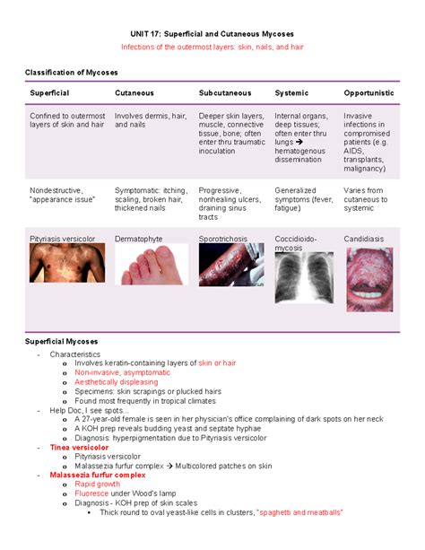
Table of Contents
- Label The Images To Review The Spectrum Of Cutaneous Mycoses
- Table of Contents
- Labeling Images to Review the Spectrum of Cutaneous Mycoses: A Comprehensive Guide
- Understanding the Spectrum of Cutaneous Mycoses
- 1. Dermatophytoses (Tineas):
- 2. Candidiasis:
- 3. Pityriasis Versicolor:
- 4. Other Cutaneous Mycoses:
- Essential Elements for Image Labeling:
- Practical Considerations for Image Analysis:
- Improving Diagnostic Accuracy:
- The Role of Image Labeling in Research and Education:
- Conclusion:
- Latest Posts
- Latest Posts
- Related Post
Labeling Images to Review the Spectrum of Cutaneous Mycoses: A Comprehensive Guide
Cutaneous mycoses, commonly known as fungal skin infections, represent a diverse group of dermatological conditions affecting millions worldwide. Accurate diagnosis hinges on a thorough clinical examination, coupled with effective image analysis and labeling. This article provides a comprehensive guide to labeling images for reviewing the spectrum of cutaneous mycoses, emphasizing key features for identification and differentiation.
Understanding the Spectrum of Cutaneous Mycoses
Cutaneous mycoses are classified based on the fungal species involved and the clinical presentation. The spectrum encompasses a wide range of infections, each with unique characteristics:
1. Dermatophytoses (Tineas):
These are caused by dermatophytes, a group of fungi that thrive in keratinized tissues like skin, hair, and nails. Common types include:
-
Tinea pedis (Athlete's foot): Characterized by scaling, maceration, and fissuring between the toes. Images should clearly depict the location, extent of involvement, and any associated inflammation or secondary bacterial infection. Keywords: Tinea pedis, athlete's foot, interdigital, scaling, maceration, fissuring.
-
Tinea cruris (Jock itch): Presents as erythematous, scaly patches in the groin area. Labeling should note the distribution, borders (often well-defined), and any satellite lesions. Keywords: Tinea cruris, jock itch, groin, erythema, scaling, satellite lesions.
-
Tinea corporis (Ringworm): Manifests as annular or circular lesions with raised, scaly borders and central clearing. Accurate labeling requires documenting lesion morphology, size, and the presence of any central clearing or scaling. Keywords: Tinea corporis, ringworm, annular, circular, scaling, central clearing.
-
Tinea capitis (Scalp ringworm): This infection affects the scalp and hair, presenting as scaly patches, alopecia, and sometimes pustules or kerions. Image labeling should focus on the location, type of hair involvement (e.g., patchy alopecia, black dots), and any inflammatory response. Keywords: Tinea capitis, scalp ringworm, alopecia, kerion, pustules, black dots.
-
Tinea unguium (Onychomycosis): This involves the nails, causing discoloration, thickening, and distortion. Labels should specify nail involvement (one or more nails), the type of changes (e.g., dystrophy, thickening, discoloration), and the extent of involvement. Keywords: Tinea unguium, onychomycosis, nail dystrophy, thickening, discoloration.
2. Candidiasis:
Caused by Candida species, typically Candida albicans, candidiasis commonly affects skin folds, mucous membranes, and nails.
-
Intertrigo: Occurs in skin folds, presenting as erythematous, macerated, and often satellite lesions. Image labels should highlight the location (e.g., axillae, inguinal folds), the presence of satellite lesions, and the degree of maceration. Keywords: Candidiasis, intertrigo, maceration, satellite lesions, erythema, skin folds.
-
Paronychia: Involves inflammation around the nails, presenting as swelling, redness, and pain. Labels should describe the extent of involvement (acute or chronic), the presence of pus, and the affected nail(s). Keywords: Candidiasis, paronychia, nail inflammation, swelling, pus.
3. Pityriasis Versicolor:
Caused by Malassezia species, this infection results in hypopigmented or hyperpigmented scaly macules on the trunk and extremities. Labels should clearly indicate lesion distribution, color changes (hypopigmented or hyperpigmented), and the presence of fine scaling. Keywords: Pityriasis versicolor, Malassezia, hypopigmented, hyperpigmented, scaling, macules.
4. Other Cutaneous Mycoses:
Less common cutaneous mycoses include:
-
Sporotrichosis: Characterized by nodular lesions along lymphatic channels. Image labeling should focus on the location of lesions, their morphology (nodular or ulcerative), and their linear arrangement along lymphatic vessels. Keywords: Sporotrichosis, nodular lesions, lymphatic channels, ulcerative lesions.
-
Chromoblastomycosis: Presents with verrucous, wart-like lesions. Labels should emphasize the verrucous nature of the lesions, their color, and any associated scarring. Keywords: Chromoblastomycosis, verrucous lesions, wart-like lesions, scarring.
Essential Elements for Image Labeling:
Effective image labeling for cutaneous mycoses requires attention to detail and consistency. The following elements are crucial:
- Patient Demographics: Age, sex, and relevant medical history.
- Lesion Location: Precise anatomical location (e.g., right foot, anterior thigh).
- Lesion Morphology: Shape, size, color, texture (e.g., annular, scaly, erythematous, papular).
- Distribution: Localized or widespread.
- Associated Findings: Satellite lesions, central clearing, inflammation, pustules, scaling, maceration, hair involvement, nail changes.
- Clinical Diagnosis (if available): Tentative diagnosis based on clinical findings.
- Date of Image Acquisition: Important for tracking disease progression.
Practical Considerations for Image Analysis:
- Image Quality: High-resolution images are essential for accurate assessment. Ensure good lighting and clear focus.
- Multiple Views: Capture images from different angles and distances to fully appreciate the lesion's characteristics.
- Magnification: Close-up images can reveal fine details such as scaling, texture, and hair involvement.
- Comparison Images: If possible, include images taken at different time points to monitor treatment response.
- Standard Nomenclature: Use consistent terminology for labeling (e.g., from standardized medical dictionaries).
Improving Diagnostic Accuracy:
Accurate labeling contributes significantly to diagnostic accuracy. Consider these points for enhancing the process:
- Correlation with Clinical Data: Always integrate the image findings with the patient's history, physical examination, and other diagnostic tests.
- Differential Diagnosis: Develop a comprehensive differential diagnosis based on the labeled image features, considering various fungal and non-fungal conditions that may mimic cutaneous mycoses.
- Consultation with Specialists: In cases of unusual or complex presentations, consultation with dermatologists or mycologists is crucial.
- Use of Dermoscopy: Dermoscopy can enhance the visualization of subtle features not readily apparent on routine examination and thus is a significant tool for improving the labeling process and accuracy.
The Role of Image Labeling in Research and Education:
Well-labeled images of cutaneous mycoses are invaluable for:
- Medical Education: Training medical students and healthcare professionals in the diagnosis and management of fungal skin infections.
- Research Studies: Creating large datasets for epidemiological studies, clinical trials, and the development of new diagnostic tools.
- Teledermatology: Facilitating remote diagnosis and management of cutaneous mycoses.
Conclusion:
Labeling images of cutaneous mycoses is a crucial step in accurate diagnosis, effective management, and advancing our understanding of these common infections. A systematic approach, emphasizing detailed descriptions of lesion characteristics and consistent terminology, is essential for maximizing the value of these images for clinical care, research, and education. By adhering to the principles outlined in this guide, healthcare professionals can significantly improve the quality of image analysis and contribute to better patient outcomes. The application of standardized labeling practices across various healthcare settings ensures consistent communication, accurate diagnoses, and effective knowledge sharing, ultimately contributing to improved global healthcare standards in the management of cutaneous mycoses. Furthermore, continued advancements in digital pathology and machine learning will only further refine this process and improve our ability to accurately diagnose and manage these often-challenging infections.
Latest Posts
Latest Posts
-
What Is The Formula For The Compound Iron Iii Sulfite
Apr 05, 2025
-
Tom Has Built A Large Slingshot
Apr 05, 2025
-
As A Hiker In Glacier National Park
Apr 05, 2025
-
3 Rs For Responding To Aggressive Behavior
Apr 05, 2025
-
A Mathematical Sentence With An Equal Symbol Used
Apr 05, 2025
Related Post
Thank you for visiting our website which covers about Label The Images To Review The Spectrum Of Cutaneous Mycoses . We hope the information provided has been useful to you. Feel free to contact us if you have any questions or need further assistance. See you next time and don't miss to bookmark.
