Label The Features Of A Neuromuscular Junction
Holbox
Mar 17, 2025 · 7 min read
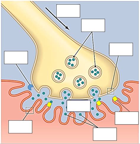
Table of Contents
Labeling the Features of a Neuromuscular Junction: A Comprehensive Guide
The neuromuscular junction (NMJ) is a specialized synapse between a motor neuron and a skeletal muscle fiber. It's the site where nerve impulses are transmitted from the neuron to the muscle, initiating muscle contraction. Understanding the intricate features of the NMJ is crucial for comprehending muscle physiology, neurological disorders, and various pharmacological interventions. This article provides a detailed exploration of the NMJ's key components, focusing on their structure and function. We will meticulously label these features to ensure a comprehensive understanding.
I. The Presynaptic Neuron: Where the Signal Begins
The presynaptic neuron, also known as the motor neuron, is the starting point of the neuromuscular transmission. Its axon terminal, upon reaching the muscle fiber, undergoes significant specialization to facilitate efficient neurotransmission.
A. Axon Terminal: The Communication Hub
The axon terminal is the bulbous ending of the motor neuron's axon. This structure is critical as it houses numerous synaptic vesicles. These vesicles are membrane-bound sacs packed with the neurotransmitter acetylcholine (ACh). The axon terminal's membrane contains voltage-gated calcium channels (Ca²⁺ channels). The influx of calcium ions (Ca²⁺) triggered by the arrival of an action potential is essential for the release of ACh into the synaptic cleft.
B. Synaptic Vesicles: Acetylcholine Storage and Release
Synaptic vesicles are spherical organelles containing a high concentration of ACh. The process of exocytosis, triggered by the calcium influx, releases ACh into the synaptic cleft. The number and size of synaptic vesicles are vital for the strength and efficiency of neuromuscular transmission. The release is a highly regulated process, ensuring precise control over muscle contraction. The vesicles themselves undergo a complex cycle of recycling and refilling with ACh.
C. Voltage-Gated Calcium Channels (Ca²⁺ Channels): The Key to Neurotransmitter Release
Voltage-gated calcium channels (Ca²⁺ channels) are transmembrane proteins embedded in the axon terminal membrane. These channels open in response to depolarization caused by the arrival of an action potential. The subsequent influx of Ca²⁺ ions triggers the fusion of synaptic vesicles with the presynaptic membrane, thereby releasing ACh into the synaptic cleft. The density and distribution of these channels are crucial for the speed and magnitude of ACh release. Dysfunction in these channels can lead to various neuromuscular disorders.
II. The Synaptic Cleft: The Communication Bridge
The synaptic cleft is the narrow gap separating the presynaptic axon terminal and the postsynaptic muscle fiber membrane. It's approximately 20-30 nm wide, a remarkably small space that allows for rapid diffusion of ACh. This space is filled with extracellular matrix proteins and enzymes that play a role in ACh degradation.
A. Acetylcholinesterase (AChE): The Cleanup Crew
Acetylcholinesterase (AChE) is a crucial enzyme located within the synaptic cleft. Its primary function is to rapidly hydrolyze ACh into choline and acetate. This hydrolysis terminates the action of ACh, preventing prolonged muscle contraction and allowing for precise control of muscle activity. The efficient action of AChE is critical for preventing muscle spasms and other neuromuscular complications. Inhibitors of AChE, such as certain insecticides and nerve gases, can lead to severe muscle paralysis due to prolonged ACh activity.
III. The Postsynaptic Muscle Fiber: The Responder
The postsynaptic membrane of the muscle fiber, also known as the motor end-plate, is highly specialized to receive and respond to the ACh released from the presynaptic neuron.
A. Motor End-Plate: The Muscle's Receiver
The motor end-plate is a specialized region of the muscle fiber membrane that lies directly opposite the axon terminal. It's characterized by a high density of acetylcholine receptors (AChRs). The motor end-plate's folded structure increases the surface area available for ACh binding, maximizing the efficiency of neurotransmission. This folded structure is crucial for the amplification of the signal. Disruptions to the motor end-plate structure can severely impair neuromuscular transmission.
B. Acetylcholine Receptors (AChRs): The Signal Transducers
Acetylcholine receptors (AChRs) are ligand-gated ion channels located on the postsynaptic membrane of the motor end-plate. These receptors bind to ACh, initiating a conformational change that opens the ion channel. This opening allows the passage of sodium ions (Na⁺) into the muscle fiber, causing depolarization of the postsynaptic membrane. This depolarization generates an end-plate potential (EPP), which is a graded potential that triggers muscle action potential. The number and functionality of AChRs are critical determinants of neuromuscular transmission efficacy. Diseases like myasthenia gravis affect AChR function, leading to muscle weakness.
C. Junctional Folds: Surface Area Maximization
The junctional folds are invaginations of the postsynaptic membrane. These folds significantly increase the surface area of the motor end-plate, thereby accommodating a higher density of AChRs. This structural feature enhances the efficiency of ACh binding and the generation of the EPP, ensuring robust neuromuscular transmission. The depth and complexity of the junctional folds vary among different muscle types and species.
D. End-Plate Potential (EPP): The Trigger for Muscle Contraction
The end-plate potential (EPP) is a graded depolarization of the postsynaptic membrane caused by the influx of Na⁺ ions through AChR channels. The EPP spreads along the muscle fiber membrane, triggering the opening of voltage-gated Na⁺ channels and the generation of a muscle action potential. The amplitude of the EPP is directly related to the amount of ACh released from the presynaptic terminal. An EPP of sufficient amplitude will consistently trigger a muscle action potential, ensuring reliable muscle contraction.
IV. Supporting Structures: Ensuring Proper Function
The NMJ is not just a collection of isolated components; it's a complex structure supported by various components that ensure its proper function and stability.
A. Basal Lamina: Structural Support and Molecular Scaffolding
The basal lamina is a thin extracellular matrix that surrounds the NMJ, providing structural support and organization. It's a complex mixture of proteins and glycoproteins that helps maintain the close apposition between the pre- and postsynaptic elements. The basal lamina plays a crucial role in the organization and maintenance of AChRs at the motor end-plate. It also contains several molecules involved in regulating NMJ development and regeneration.
B. Schwann Cells: Myelinating Support
Although primarily associated with myelinated axons, Schwann cells also play a role in the NMJ. They contribute to the formation and maintenance of the basal lamina and participate in the trophic support of the axon terminal. Schwann cells are not directly involved in neurotransmission but provide essential structural support to the NMJ.
V. Clinical Significance: Understanding Neuromuscular Disorders
Understanding the NMJ’s intricate features is crucial for diagnosing and treating various neuromuscular disorders. Disruptions at any level of the NMJ can result in muscle weakness, paralysis, or other debilitating conditions.
A. Myasthenia Gravis: Autoimmune Attack on AChRs
Myasthenia gravis is an autoimmune disorder characterized by the production of antibodies against AChRs. This results in a reduction in the number of functional AChRs at the motor end-plate, leading to muscle weakness and fatigue. Treatment strategies often focus on increasing ACh levels or inhibiting AChE to enhance neuromuscular transmission.
B. Lambert-Eaton Myasthenic Syndrome (LEMS): Presynaptic Dysfunction
Lambert-Eaton myasthenic syndrome (LEMS) is a less common disorder affecting the presynaptic neuron. Antibodies target voltage-gated Ca²⁺ channels at the axon terminal, reducing the amount of ACh released. This leads to muscle weakness, particularly in proximal muscles. Treatment strategies often involve Ca²⁺ channel modulators or immunosuppressants.
C. Botulism: Presynaptic Blockade
Botulism, caused by the toxin produced by Clostridium botulinum, blocks the release of ACh at the presynaptic terminal. This results in flaccid paralysis, as muscle contraction is severely impaired. Ironically, botulinum toxin is used therapeutically in small doses to treat certain muscle spasms and other neurological conditions.
This comprehensive exploration of the neuromuscular junction highlights its complexity and vital role in muscle function. The intricate interplay between the presynaptic neuron, the synaptic cleft, and the postsynaptic muscle fiber underscores the importance of precise regulation in neuromuscular transmission. Understanding the various components and their interactions provides a robust foundation for comprehending normal muscle physiology and the pathophysiology of various neuromuscular disorders. Furthermore, a thorough grasp of the NMJ is essential for developing targeted therapeutic interventions for these conditions.
Latest Posts
Latest Posts
-
Meiosis Starts With A Single Diploid Cell And Produces
Mar 18, 2025
-
Did The Precipitated Agcl Dissolve Explain
Mar 18, 2025
-
Which Of The Following Documents Are Considered A Record
Mar 18, 2025
-
Chegg Tinder Gold Code Not Working
Mar 18, 2025
-
Supply Creates Its Own Demand Arrows
Mar 18, 2025
Related Post
Thank you for visiting our website which covers about Label The Features Of A Neuromuscular Junction . We hope the information provided has been useful to you. Feel free to contact us if you have any questions or need further assistance. See you next time and don't miss to bookmark.
