Label The Components Of A Synapse
Holbox
Mar 16, 2025 · 7 min read
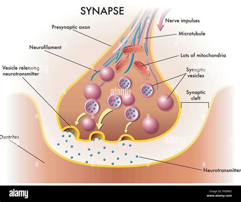
Table of Contents
Labeling the Components of a Synapse: A Deep Dive into Neuronal Communication
The synapse, the fundamental unit of communication in the nervous system, is a fascinating and complex structure. Understanding its components is crucial to grasping the intricacies of brain function, learning, and various neurological disorders. This comprehensive guide will delve into the detailed anatomy of a synapse, labeling each component and explaining its function in the intricate process of neuronal signaling. We'll explore both chemical and electrical synapses, highlighting their similarities and key differences. By the end, you'll have a robust understanding of this vital structure.
Types of Synapses: Chemical vs. Electrical
Before diving into the specific components, it's important to establish the two main types of synapses:
Chemical Synapses: The Majority
The vast majority of synapses in the nervous system are chemical synapses. These synapses rely on the release of neurotransmitters, chemical messengers, to transmit signals from the presynaptic neuron to the postsynaptic neuron. This process involves a gap, called the synaptic cleft, which prevents direct electrical transmission.
Electrical Synapses: Direct Transmission
Electrical synapses, on the other hand, allow for the direct flow of electrical current between neurons through specialized protein channels called gap junctions. These junctions create a direct physical connection between the pre- and postsynaptic neurons, enabling rapid and synchronized transmission of signals. While faster, they are less flexible in terms of signal modulation than chemical synapses.
Components of a Chemical Synapse: A Detailed Look
Let's now focus on the intricate components of a chemical synapse, the more prevalent type:
1. Presynaptic Neuron: The Sender
The presynaptic neuron is the neuron sending the signal. Key components within the presynaptic terminal include:
-
Synaptic Vesicles: These small, membrane-bound sacs store and release neurotransmitters. They are crucial for the chemical transmission process. The number and size of vesicles can vary depending on the type of synapse and the activity level of the neuron.
-
Neurotransmitters: These chemical messengers are synthesized within the presynaptic neuron and packaged into synaptic vesicles. Examples include acetylcholine, dopamine, serotonin, GABA, and glutamate. Each neurotransmitter has a specific effect on the postsynaptic neuron, influencing its excitability or inhibitory activity.
-
Voltage-Gated Calcium Channels: These channels are essential for neurotransmitter release. When an action potential reaches the presynaptic terminal, these channels open, allowing calcium ions (Ca²⁺) to enter the terminal. The influx of Ca²⁺ triggers the fusion of synaptic vesicles with the presynaptic membrane, releasing neurotransmitters into the synaptic cleft.
-
Presynaptic Membrane: This is the membrane of the presynaptic terminal, the site where neurotransmitters are released. It's highly specialized with proteins involved in vesicle fusion and recycling.
2. Synaptic Cleft: The Gap
The synaptic cleft is a narrow space (approximately 20-40 nm) separating the presynaptic and postsynaptic membranes. This gap prevents direct electrical transmission and ensures that the signal transmission is chemically mediated. The cleft's composition includes extracellular matrix molecules that help maintain the structural integrity of the synapse and regulate the diffusion of neurotransmitters.
3. Postsynaptic Neuron: The Receiver
The postsynaptic neuron receives the signal transmitted by the presynaptic neuron. Key components include:
-
Postsynaptic Membrane: This membrane contains receptor proteins specific to the neurotransmitter released by the presynaptic neuron. The binding of neurotransmitters to these receptors initiates a response in the postsynaptic neuron.
-
Receptors: These specialized proteins are embedded in the postsynaptic membrane. They bind to specific neurotransmitters, initiating a cascade of events that alter the postsynaptic neuron's membrane potential. Receptors can be either ionotropic (directly opening ion channels) or metabotropic (indirectly influencing ion channels through intracellular signaling pathways).
-
Postsynaptic Density (PSD): This electron-dense region on the postsynaptic membrane is a complex scaffold of proteins that anchors receptors, signaling molecules, and other proteins involved in synaptic plasticity and signal transduction. It's a crucial area for the integration of synaptic signals and the modulation of synaptic strength.
-
Ion Channels: These channels allow the passage of specific ions across the postsynaptic membrane. The opening or closing of these channels, triggered by receptor activation, alters the membrane potential of the postsynaptic neuron, leading to either excitation or inhibition.
4. Supporting Cells: Glia
While not directly part of the synaptic structure, glial cells, such as astrocytes and oligodendrocytes (in the CNS) and Schwann cells (in the PNS), play crucial supportive roles.
-
Astrocytes: These cells regulate the extracellular environment around the synapse, influencing neurotransmitter uptake and recycling. They also help maintain the structural integrity of the synapse.
-
Oligodendrocytes/Schwann cells: These cells produce myelin, an insulating sheath that surrounds axons, facilitating faster and more efficient signal transmission.
Components of an Electrical Synapse: A Simpler Structure
Electrical synapses are structurally simpler than chemical synapses. Their key components include:
-
Gap Junctions: These are channels formed by the connection of connexons, which are hemichannels from the pre- and postsynaptic neurons. They directly link the cytoplasm of the two neurons, allowing for the direct passage of ions and small molecules.
-
Connexons: Each connexon is composed of six connexin proteins. These proteins form a pore that allows for the direct passage of ions and small molecules between the two connected neurons.
Synaptic Transmission: The Process
Understanding the components is only half the battle. The process of synaptic transmission itself is crucial to grasp:
Chemical Synapse Transmission:
-
Action Potential Arrival: An action potential reaches the presynaptic terminal.
-
Calcium Influx: Voltage-gated calcium channels open, allowing calcium ions to enter the terminal.
-
Vesicle Fusion: Calcium influx triggers the fusion of synaptic vesicles with the presynaptic membrane.
-
Neurotransmitter Release: Neurotransmitters are released into the synaptic cleft.
-
Neurotransmitter Binding: Neurotransmitters diffuse across the cleft and bind to receptors on the postsynaptic membrane.
-
Postsynaptic Response: Receptor binding triggers a change in the postsynaptic membrane potential, either excitatory (depolarization) or inhibitory (hyperpolarization).
-
Neurotransmitter Removal: Neurotransmitters are removed from the synaptic cleft through reuptake, enzymatic degradation, or diffusion.
Electrical Synapse Transmission:
-
Action Potential Arrival: An action potential reaches the presynaptic neuron.
-
Direct Current Flow: The electrical current flows directly through the gap junctions from the presynaptic to the postsynaptic neuron.
-
Postsynaptic Response: The direct current flow causes a change in the postsynaptic membrane potential.
This simplified explanation highlights the core steps; the intricacies involve numerous regulatory proteins and signaling pathways.
Clinical Relevance: Synaptic Dysfunction and Neurological Disorders
Dysfunction in synaptic transmission is implicated in a wide range of neurological and psychiatric disorders, including:
-
Alzheimer's disease: Characterized by impaired synaptic plasticity and neurotransmitter dysfunction.
-
Parkinson's disease: Involves a deficiency in dopamine neurotransmission in the brain.
-
Epilepsy: Often associated with imbalances in excitation and inhibition in neuronal circuits.
-
Schizophrenia: Linked to disruptions in dopamine and glutamate neurotransmission.
-
Depression: Implicated in dysfunction of various neurotransmitter systems, including serotonin and norepinephrine.
Understanding the intricate components of the synapse and the mechanisms of synaptic transmission is essential for developing effective treatments for these and other neurological disorders. Further research continues to unravel the complexities of this vital structure and its role in brain function.
Conclusion: A Crucial Understanding
This detailed exploration of synaptic components underscores the remarkable complexity of neuronal communication. From the presynaptic vesicles brimming with neurotransmitters to the postsynaptic receptors meticulously awaiting their arrival, every component plays a critical role in the seamless transmission of information throughout the nervous system. The intricate dance between chemical and electrical signals, mediated by a myriad of proteins and supported by glial cells, forms the very basis of our thoughts, feelings, and actions. A deep understanding of synaptic function is not merely an academic pursuit but a vital step toward deciphering the mysteries of the brain and developing effective therapies for neurological diseases. Further study into the specific types of neurotransmitters, receptor subtypes, and the modulatory effects of various factors will continue to enrich our understanding of this fundamental aspect of neuroscience.
Latest Posts
Latest Posts
-
Balance The Following Equations By Inserting Coefficients As Needed
Mar 16, 2025
-
Prepaid Accounts Also Called Prepaid Expenses Are
Mar 16, 2025
-
Fermentation In Yeast Can Occur Without
Mar 16, 2025
-
What Is Hootsuite Inbox Is Used For Pick Three
Mar 16, 2025
-
Which Distribution Channel Drives Results Fastest
Mar 16, 2025
Related Post
Thank you for visiting our website which covers about Label The Components Of A Synapse . We hope the information provided has been useful to you. Feel free to contact us if you have any questions or need further assistance. See you next time and don't miss to bookmark.
