Label The Bony Structures Of The Shoulder And Upper Limb.
Holbox
Mar 24, 2025 · 6 min read
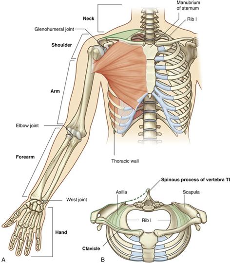
Table of Contents
- Label The Bony Structures Of The Shoulder And Upper Limb.
- Table of Contents
- Labeling the Bony Structures of the Shoulder and Upper Limb: A Comprehensive Guide
- The Shoulder Girdle: Clavicle and Scapula
- The Clavicle: The Collarbone's Key Features
- The Scapula: The Shoulder Blade's Complex Anatomy
- The Upper Limb: Humerus, Radius, Ulna, Carpals, Metacarpals, and Phalanges
- The Humerus: The Arm Bone
- The Radius and Ulna: The Forearm Bones
- The Hand Bones: Carpals, Metacarpals, and Phalanges
- Clinical Significance
- Conclusion
- Latest Posts
- Latest Posts
- Related Post
Labeling the Bony Structures of the Shoulder and Upper Limb: A Comprehensive Guide
The shoulder and upper limb, a marvel of human biomechanics, boasts a complex arrangement of bones, joints, and muscles that allow for a remarkable range of motion and dexterity. Understanding the bony anatomy of this region is crucial for healthcare professionals, students of anatomy, and anyone interested in human movement and physiology. This guide provides a comprehensive overview of the bony structures, their articulations, and key landmarks, focusing on accurate labeling and understanding their spatial relationships. We'll delve into each bone in detail, exploring their unique features and clinical significance.
The Shoulder Girdle: Clavicle and Scapula
The shoulder girdle, the foundation upon which the upper limb rests, consists of two bones: the clavicle (collarbone) and the scapula (shoulder blade). These bones articulate with each other and with the sternum and humerus, forming a dynamic and mobile structure.
The Clavicle: The Collarbone's Key Features
The clavicle, a long bone with a distinctive S-shape, is easily palpable along the superior anterior aspect of the thorax. Its medial end articulates with the manubrium of the sternum at the sternoclavicular joint (SC joint), the only bony connection between the upper limb and the axial skeleton. The lateral end articulates with the acromion process of the scapula at the acromioclavicular joint (AC joint).
Key Landmarks of the Clavicle:
- Sternal End: The medial, rounded end articulating with the sternum.
- Acromial End: The flattened lateral end articulating with the acromion.
- Conoid Tubercle: A roughened area on the inferior surface, near the acromial end, for ligament attachment.
- Costal Groove: A shallow groove on the inferior surface, running along most of its length.
The Scapula: The Shoulder Blade's Complex Anatomy
The scapula, a flat, triangular bone, sits on the posterior aspect of the thorax, overlying ribs 2-7. Its remarkable mobility allows for a wide range of shoulder movements.
Key Landmarks of the Scapula:
- Spine: A prominent ridge running across the posterior surface, ending in the acromion.
- Acromion: A lateral extension of the spine, articulating with the clavicle.
- Coracoid Process: A hook-like projection extending anteriorly from the superior border. This serves as an attachment site for several important muscles.
- Glenoid Cavity: A shallow, pear-shaped fossa on the lateral aspect, articulating with the head of the humerus to form the glenohumeral joint.
- Suprascapular Notch: A notch on the superior border, often converted into a foramen by a transverse ligament.
- Superior Border: The superior margin of the scapula.
- Medial (Vertebral) Border: The medial margin of the scapula, parallel to the vertebral column.
- Lateral (Axillary) Border: The lateral margin of the scapula, facing towards the axilla.
- Inferior Angle: The inferior and lateral point of the scapula.
- Superior Angle: The superior and medial point of the scapula.
The Upper Limb: Humerus, Radius, Ulna, Carpals, Metacarpals, and Phalanges
The free upper limb, extending from the shoulder girdle to the fingertips, comprises several bones that work together to provide incredible dexterity and mobility.
The Humerus: The Arm Bone
The humerus, the longest bone in the upper limb, consists of a proximal head, a shaft, and a distal end. It articulates with the scapula proximally at the glenohumeral joint and with the radius and ulna distally at the elbow joint.
Key Landmarks of the Humerus:
- Head: The rounded proximal end, articulating with the glenoid cavity of the scapula.
- Anatomical Neck: A constricted area just distal to the head.
- Greater Tubercle: A large projection on the lateral aspect, serving as an attachment site for muscles.
- Lesser Tubercle: A smaller projection on the anterior medial aspect, also serving as a muscle attachment site.
- Intertubercular Sulcus (Bicipital Groove): A groove between the greater and lesser tubercles, containing the tendon of the long head of the biceps brachii.
- Surgical Neck: A constricted area distal to the tubercles, a common site for fractures.
- Deltoid Tuberosity: A roughened area on the lateral aspect, for the insertion of the deltoid muscle.
- Radial Groove: A groove on the posterior surface, running distally, containing the radial nerve.
- Capitulum: A rounded articular surface on the lateral aspect of the distal end, articulating with the head of the radius.
- Trochlea: A spool-shaped articular surface on the medial aspect of the distal end, articulating with the trochlear notch of the ulna.
- Medial Epicondyle: A bony prominence on the medial aspect of the distal end.
- Lateral Epicondyle: A bony prominence on the lateral aspect of the distal end.
- Olecranon Fossa: A deep depression on the posterior aspect of the distal end, receiving the olecranon process of the ulna during elbow extension.
- Coronoid Fossa: A shallow depression on the anterior aspect of the distal end, receiving the coronoid process of the ulna during elbow flexion.
The Radius and Ulna: The Forearm Bones
The radius and ulna are two long bones that form the forearm. They articulate with each other proximally and distally, allowing for pronation and supination (rotation of the forearm). The radius articulates with the humerus, and the ulna primarily articulates with the humerus, while both articulate with the carpals.
Key Landmarks of the Radius:
- Head: The disc-shaped proximal end, articulating with the capitulum of the humerus and the radial notch of the ulna.
- Radial Tuberosity: A roughened area on the medial aspect, for the insertion of the biceps brachii muscle.
- Styloid Process: A pointed projection on the lateral aspect of the distal end.
Key Landmarks of the Ulna:
- Olecranon Process: The large, hook-like projection forming the point of the elbow.
- Trochlear Notch: A curved articular surface that receives the trochlea of the humerus.
- Coronoid Process: A projection anterior to the trochlear notch.
- Radial Notch: A shallow depression on the lateral side of the proximal end that receives the head of the radius.
- Styloid Process: A pointed projection on the medial aspect of the distal end.
The Hand Bones: Carpals, Metacarpals, and Phalanges
The hand is composed of three groups of bones: the carpals, metacarpals, and phalanges.
- Carpals: Eight small, irregularly shaped bones arranged in two rows. These bones form the wrist and allow for complex movements. They are named: Scaphoid, Lunate, Triquetrum, Pisiform, Trapezium, Trapezoid, Capitate, Hamate.
- Metacarpals: Five long bones forming the palm of the hand. They are numbered I-V, starting from the thumb side.
- Phalanges: Fourteen bones forming the fingers. Each finger (except the thumb) has three phalanges: proximal, middle, and distal. The thumb has only two: proximal and distal.
Clinical Significance
A thorough understanding of the bony landmarks of the shoulder and upper limb is critical for diagnosing and managing various clinical conditions. Fractures, dislocations, and sprains are common injuries in this region. Accurate labeling and identification of bony structures are essential for:
- Imaging Interpretation: Radiographs, CT scans, and MRIs rely on the accurate identification of bony landmarks for diagnosis.
- Surgical Planning: Precise knowledge of bone anatomy is crucial for surgical procedures involving the shoulder and upper limb.
- Neurological Assessment: The bony structures provide anatomical reference points for assessing nerve function and detecting potential nerve compression.
- Palpation and Physical Examination: Identifying bony landmarks assists in performing a thorough physical examination and assessing the range of motion.
Conclusion
Labeling the bony structures of the shoulder and upper limb requires a systematic approach combining anatomical knowledge with careful observation and practical experience. This detailed guide provides a comprehensive overview of each bone, highlighting key landmarks and their clinical significance. Remember that consistent practice and hands-on learning are key to mastering this intricate anatomical region. By thoroughly understanding the bones and their articulations, you’ll gain a deeper appreciation for the remarkable functionality and resilience of the human shoulder and upper limb. Further exploration using anatomical models, atlases, and real-world clinical observations will significantly enhance your understanding and contribute to a more profound appreciation of human anatomy.
Latest Posts
Latest Posts
-
Which Of The Following Statements Are Correct
Mar 26, 2025
-
What Is Shown In The Image
Mar 26, 2025
-
Draw The Product S Of The Following Reactions
Mar 26, 2025
-
Which Of The Following Is Not A Property Of Water
Mar 26, 2025
-
Which Of The Following Statements About Platform Businesses Is True
Mar 26, 2025
Related Post
Thank you for visiting our website which covers about Label The Bony Structures Of The Shoulder And Upper Limb. . We hope the information provided has been useful to you. Feel free to contact us if you have any questions or need further assistance. See you next time and don't miss to bookmark.
