Label The Arteries Of The Right Lower Limb.
Holbox
Mar 23, 2025 · 5 min read
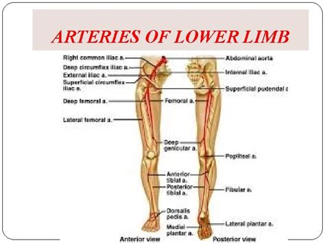
Table of Contents
- Label The Arteries Of The Right Lower Limb.
- Table of Contents
- Labeling the Arteries of the Right Lower Limb: A Comprehensive Guide
- The Arterial Supply: Beginning at the Iliac Arteries
- The Femoral Artery and its Branches
- Transition to the Popliteal Artery: Behind the Knee
- Distal Branches: The Tibial Arteries and their Branches
- The Anterior Tibial Artery: To the Front of the Leg
- The Posterior Tibial Artery: To the Back of the Leg
- Clinical Significance and Conclusion
- Latest Posts
- Latest Posts
- Related Post
Labeling the Arteries of the Right Lower Limb: A Comprehensive Guide
The arteries of the lower limb form a complex network responsible for delivering oxygenated blood to the muscles, bones, and tissues of the leg and foot. Understanding their branching patterns and locations is crucial for medical professionals, anatomy students, and anyone interested in the intricacies of the human circulatory system. This comprehensive guide will walk you through the arteries of the right lower limb, providing detailed descriptions and visual aids (although images themselves are not included in this text-based format). We'll use a systematic approach, moving from the proximal (closest to the body) to the distal (farthest from the body) segments.
The Arterial Supply: Beginning at the Iliac Arteries
The arterial supply to the right lower limb originates from the external iliac artery, which continues distally as the femoral artery after passing under the inguinal ligament. This is a critical landmark in understanding the lower limb's vascular anatomy.
The Femoral Artery and its Branches
The femoral artery, a large and significant vessel, runs down the anterior thigh. It gives off several important branches:
- Superficial Epigastric Artery: A small branch supplying the lower abdominal wall.
- Superficial Circumflex Iliac Artery: Another small branch supplying the anterior superior iliac spine and surrounding area.
- Superficial External Pudendal Artery: This artery provides blood to the external genitalia. It's important to note its superficial position.
- Deep External Pudendal Artery: A deeper branch compared to its superficial counterpart, also contributing to the blood supply of the external genitalia.
- Deep Femoral Artery (Profunda Femoris Artery): This is a substantially larger branch of the femoral artery. It runs posteriorly and supplies most of the deep muscles of the thigh. The profunda femoris artery itself gives off several crucial branches:
- Medial Circumflex Femoral Artery: This branch wraps around the femur, supplying blood to the head and neck of the femur. It's crucial for femoral head survival.
- Lateral Circumflex Femoral Artery: This artery also circles the femur, contributing to the blood supply of the femur and surrounding muscles.
- Perforating Arteries: Several perforating arteries pierce the adductor magnus muscle, supplying blood to the posterior compartment of the thigh.
Transition to the Popliteal Artery: Behind the Knee
As the femoral artery passes through the adductor hiatus (an opening in the adductor magnus muscle), it enters the popliteal fossa (the hollow behind the knee) and becomes the popliteal artery. This is another pivotal point in the arterial pathway.
The popliteal artery is relatively short but crucial. It gives rise to several branches that supply the knee joint and surrounding structures. These branches include:
- Genicular Arteries: These are multiple small arteries supplying the knee joint itself. Superior and inferior medial and lateral genicular arteries ensure comprehensive blood supply to this complex joint. Their importance in knee health cannot be overstated.
Distal Branches: The Tibial Arteries and their Branches
The popliteal artery then bifurcates (splits into two) at the lower border of the popliteus muscle, giving rise to the anterior tibial artery and the posterior tibial artery.
The Anterior Tibial Artery: To the Front of the Leg
The anterior tibial artery travels down the anterior compartment of the leg, passing between the tibia and fibula. It's named for its anterior location relative to the tibia. Along its course, it gives off smaller branches to the anterior leg muscles.
As it approaches the ankle joint, the anterior tibial artery becomes the dorsalis pedis artery, a crucial vessel on the dorsum (top) of the foot. The dorsalis pedis artery is palpable and often used to assess peripheral circulation. Its branches contribute to the arterial supply of the foot and toes. It gives off branches such as:
- Arcuate Artery: This artery forms an arch across the metatarsals.
- Dorsal Metatarsal Arteries: Branching from the arcuate artery, these supply the metatarsals.
- Digital Arteries: These are the terminal branches, supplying the toes themselves.
The Posterior Tibial Artery: To the Back of the Leg
The posterior tibial artery descends down the posterior compartment of the leg, deep to the muscles of the calf. This artery is essential for the blood supply to the calf muscles.
Along its path, the posterior tibial artery gives off several important branches:
- Peroneal Artery (Fibular Artery): A substantial branch that runs along the fibula, supplying the lateral compartment of the leg.
- Medial and Lateral Plantar Arteries: These arteries originate at the ankle and provide blood to the plantar (sole) aspect of the foot. They are critical for the function and health of the foot. The plantar arteries are deeply positioned, unlike the dorsalis pedis. They also contribute to the formation of plantar arches.
Clinical Significance and Conclusion
Understanding the arterial anatomy of the right lower limb is crucial for several reasons:
-
Diagnosis and Treatment of Peripheral Artery Disease (PAD): PAD is a condition affecting blood flow in the arteries. Knowing the arterial pathways is vital for diagnosis and intervention. Palpating pulses (like the dorsalis pedis and posterior tibial pulses) is a key clinical examination technique.
-
Surgical Procedures: Vascular surgeons rely on detailed knowledge of the arterial anatomy to plan and execute procedures such as angioplasty, bypass surgery, and endarterectomy.
-
Trauma Management: In cases of trauma to the lower limb, understanding the arterial supply is critical for managing blood loss and ensuring adequate perfusion to the tissues.
-
Medical Imaging Interpretation: Radiologists and other medical professionals utilize imaging techniques such as angiography to visualize the arteries. Accurate interpretation requires a thorough understanding of normal arterial anatomy.
In conclusion, the arterial supply to the right lower limb is a complex but fascinating system. From the iliac arteries to the digital arteries of the toes, each branch plays a vital role in delivering oxygenated blood to the tissues. This detailed overview highlights the key arteries and their branches, emphasizing the clinical importance of understanding this intricate vascular network. Further study using anatomical models, diagrams, and cadaveric dissections is recommended for a deeper understanding of the three-dimensional relationships and variations that can occur in individual anatomy. Remember that this information is for educational purposes and should not be substituted for professional medical advice. Always consult a qualified healthcare professional for any health concerns.
Latest Posts
Latest Posts
-
In Worldview What Is Human Nature
Mar 25, 2025
-
How Many Moles Of K2so4 Are In 15 0g Of K2so4
Mar 25, 2025
-
Which Polymers Are Composed Of Amino Acids
Mar 25, 2025
-
Varcarolis Foundations Of Psychiatric Mental Health Nursing
Mar 25, 2025
-
A Baseball Player Is Sliding Into Second Base
Mar 25, 2025
Related Post
Thank you for visiting our website which covers about Label The Arteries Of The Right Lower Limb. . We hope the information provided has been useful to you. Feel free to contact us if you have any questions or need further assistance. See you next time and don't miss to bookmark.
