How Is Magnification Controlled In A Microscope
Holbox
Apr 07, 2025 · 6 min read
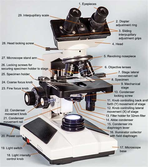
Table of Contents
- How Is Magnification Controlled In A Microscope
- Table of Contents
- How is Magnification Controlled in a Microscope? A Deep Dive into Optical and Digital Zoom
- Understanding Magnification: A Foundation
- Optical Magnification: The Heart of Microscopy
- Objective Lenses: The Key Players
- Eyepieces (Ocular Lenses): Refining the View
- Digital Magnification: Enhancing and Extending Capabilities
- Advanced Magnification Techniques: Pushing the Boundaries
- Factors Affecting Magnification Quality
- Best Practices for Magnification Control
- Conclusion: Mastering the Art of Magnification
- Latest Posts
- Latest Posts
- Related Post
How is Magnification Controlled in a Microscope? A Deep Dive into Optical and Digital Zoom
Microscopes, the indispensable tools of scientific exploration, allow us to visualize the intricate details of the microscopic world, from the cellular structures of living organisms to the crystalline formations of minerals. But how exactly do these instruments achieve such remarkable magnification? Understanding magnification control is key to mastering microscopy, and this comprehensive guide will delve into the intricacies of both optical and digital magnification, exploring the mechanisms, limitations, and best practices for each.
Understanding Magnification: A Foundation
Before delving into the specifics of control, let's establish a clear understanding of magnification itself. Magnification refers to the process of enlarging an image, making it appear larger than it actually is. In microscopy, this is achieved through a combination of lenses, each contributing to the overall magnification power. The total magnification is calculated by multiplying the magnification of the objective lens by the magnification of the eyepiece (ocular) lens.
Formula: Total Magnification = Objective Lens Magnification x Eyepiece Lens Magnification
For example, a 10x objective lens combined with a 10x eyepiece lens provides a total magnification of 100x. This means the image appears 100 times larger than its actual size.
Optical Magnification: The Heart of Microscopy
Optical magnification is the primary method used in most microscopes. It relies on the principles of refraction and the careful arrangement of lenses to bend and focus light, creating a magnified image. The control of optical magnification involves manipulating the objective lens, which is the lens closest to the specimen.
Objective Lenses: The Key Players
Objective lenses are the workhorses of optical magnification. They come in a variety of magnifications, typically ranging from 4x (low power) to 100x (oil immersion high power) and even higher in specialized microscopes. These lenses are precisely designed with multiple lens elements to correct for aberrations and deliver a clear, sharp image.
Types of Objective Lenses:
- Achromatic lenses: Correct for chromatic aberration (color fringing) for two wavelengths of light, usually red and blue.
- Apochromatic lenses: Correct for chromatic aberration for more wavelengths and also reduce spherical aberration (blurring due to lens curvature).
- Plan lenses: Further reduce field curvature, ensuring that the entire field of view is in sharp focus.
Controlling Objective Lenses:
The selection of the objective lens directly controls the optical magnification. Most microscopes have a rotating nosepiece (turret) that allows the user to easily switch between different objective lenses. Simply rotating the nosepiece into position brings the selected objective lens into the optical path, changing the magnification. This simple yet effective mechanism is the fundamental method of controlling optical magnification.
Eyepieces (Ocular Lenses): Refining the View
While objective lenses provide the bulk of the magnification, eyepieces further magnify the image produced by the objective. Eyepieces typically have a magnification of 10x, but other magnifications are available. The eyepiece magnification is a relatively fixed factor, unlike the easily selectable objective lenses. However, choosing eyepieces with different magnifications can subtly affect the overall magnification and field of view.
Digital Magnification: Enhancing and Extending Capabilities
Digital magnification, unlike optical magnification, does not increase the resolution of the image. It simply enlarges the existing digital image captured by a microscope camera. This is done through software processing, digitally interpolating the pixels to make the image appear larger. While useful for presentation or detailed examination of a specific area, it doesn't provide the same detail as increasing the optical magnification.
How Digital Magnification Works:
A digital camera or sensor is integrated into the microscope's system. The image captured by the sensor is then processed by software. Digital zoom functionality within the software allows the user to "zoom in" on the digital image, effectively increasing its size.
Limitations of Digital Magnification:
It is crucial to understand that digital magnification does not enhance resolution. Enlarging a digital image beyond its native resolution results in pixelation, a loss of detail, and a reduction in image quality. Therefore, it's essential to rely primarily on optical magnification for optimal detail and resolution.
Advanced Magnification Techniques: Pushing the Boundaries
Beyond the basic principles of optical and digital magnification, several advanced techniques allow researchers to achieve even greater magnification and resolve finer details.
Oil Immersion:
The 100x objective lens often employs oil immersion. A drop of immersion oil is placed between the lens and the coverslip. This oil has a refractive index similar to glass, minimizing light refraction and maximizing light transmission to the objective lens, resulting in improved resolution and clarity.
Confocal Microscopy:
Confocal microscopy uses a pinhole aperture to eliminate out-of-focus light, significantly improving the resolution and clarity of images, especially in thick samples. This technique allows for high-resolution three-dimensional imaging.
Electron Microscopy:
Electron microscopy utilizes a beam of electrons instead of light to illuminate the sample. The much shorter wavelength of electrons allows for significantly higher resolution than even the most powerful optical microscopes. This technique is crucial for imaging structures at the nanoscale.
Factors Affecting Magnification Quality
Beyond the basic controls, various factors influence the quality of the magnified image.
Resolution: Resolution refers to the ability to distinguish between two closely spaced points. Higher resolution means finer details can be observed. Optical resolution is limited by the wavelength of light used.
Aberrations: Lens imperfections, such as chromatic aberration (color fringing) and spherical aberration (blurring due to lens curvature), can degrade image quality. High-quality objective lenses are designed to minimize these aberrations.
Specimen Preparation: Proper preparation of the specimen is crucial for obtaining a clear and informative image. This includes techniques like staining, sectioning, and mounting.
Lighting: Adequate and correctly adjusted lighting is essential. Too much or too little light can negatively impact the image quality. Koehler illumination is a technique used to optimize the lighting for microscopy.
Best Practices for Magnification Control
- Start with low magnification: Begin with the lowest magnification objective (usually 4x) to locate the area of interest. Gradually increase magnification as needed.
- Adjust focus carefully: Always ensure the specimen is in sharp focus at each magnification level. Fine focus adjustments are crucial, especially at higher magnifications.
- Understand the limitations of digital zoom: Use digital zoom sparingly and only to enhance the viewing of a section already well resolved at optical magnification.
- Maintain proper illumination: Ensure adequate and even illumination throughout the magnification range.
- Regular cleaning: Keep lenses clean to maintain optimal image quality.
Conclusion: Mastering the Art of Magnification
Controlling magnification in a microscope involves a delicate balance between optical and digital techniques. While the selection of objective lenses directly influences optical magnification, digital zoom offers supplemental enlargement. However, it is crucial to understand the limitations of digital magnification, especially in relation to resolution. By mastering the principles and best practices outlined in this guide, microscopists can harness the power of magnification to explore the wonders of the microscopic world with precision and clarity. Understanding the interplay between objective lenses, eyepieces, and digital enhancement is crucial for achieving optimal image quality and extracting the maximum information from any sample.
Latest Posts
Latest Posts
-
The Heart Is Blank To The Lungs
Apr 14, 2025
-
Maternal Newborn Online Practice 2023 A
Apr 14, 2025
-
Which Of The Following Can You Expect From Opportunity Teams
Apr 14, 2025
-
Ethylene Oxide Is Produced By The Catalytic Oxidation
Apr 14, 2025
-
3z 5e 4 Methyl 3 5 Nonadiene
Apr 14, 2025
Related Post
Thank you for visiting our website which covers about How Is Magnification Controlled In A Microscope . We hope the information provided has been useful to you. Feel free to contact us if you have any questions or need further assistance. See you next time and don't miss to bookmark.