Correctly Label The Posterior Muscles Of The Thigh
Holbox
Apr 01, 2025 · 6 min read
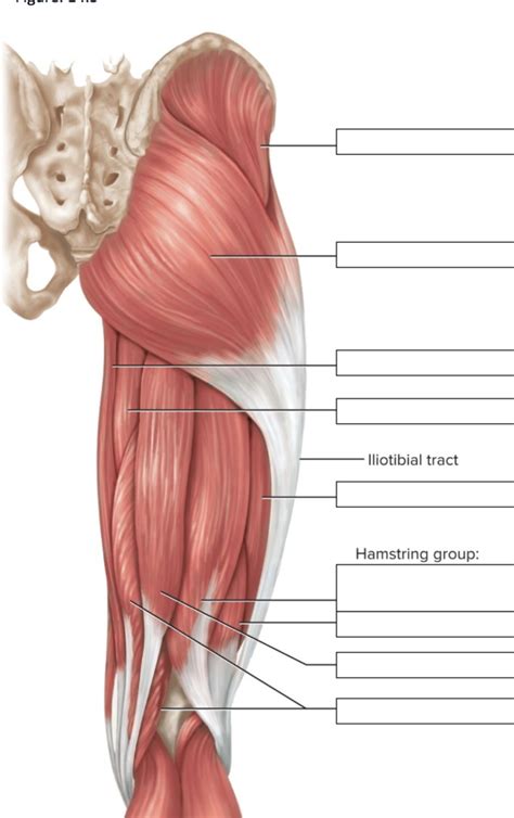
Table of Contents
- Correctly Label The Posterior Muscles Of The Thigh
- Table of Contents
- Correctly Labeling the Posterior Muscles of the Thigh: A Comprehensive Guide
- Understanding the Hamstring Muscle Group
- 1. Biceps Femoris: The Lateral Powerhouse
- 2. Semitendinosus: The Long and Slender Muscle
- 3. Semimembranosus: The Deep and Broad Muscle
- Differentiating the Hamstring Muscles: Key Anatomical Features
- Clinical Significance and Potential for Confusion
- Practical Tips for Accurate Labeling
- Beyond the Three Main Muscles: Accessory Muscles
- Conclusion: Mastering Posterior Thigh Muscle Labeling
- Latest Posts
- Latest Posts
- Related Post
Correctly Labeling the Posterior Muscles of the Thigh: A Comprehensive Guide
The posterior thigh, also known as the hamstring region, is a complex area containing several muscles crucial for hip extension and knee flexion. Accurate labeling of these muscles is essential for anyone studying anatomy, physical therapy, athletic training, or related fields. This comprehensive guide will delve into the detailed anatomy of the posterior thigh muscles, providing clear descriptions, visual aids (though not actual images due to limitations of this text-based format), and practical tips for accurate identification.
Understanding the Hamstring Muscle Group
The hamstring group is comprised of three main muscles: the biceps femoris, semitendinosus, and semimembranosus. While often grouped together, each muscle has unique characteristics regarding its origin, insertion, action, and innervation. Understanding these individual nuances is key to accurate labeling.
1. Biceps Femoris: The Lateral Powerhouse
The biceps femoris is the most lateral of the hamstring muscles. It’s unique in having two heads:
- Long head: Originates from the ischial tuberosity (the bony prominence you sit on). This shared origin with the semitendinosus and semimembranosus highlights their functional relationship.
- Short head: Originates from the linea aspera (a roughened line on the posterior femur) and the lateral supracondylar line of the femur. This distinction sets it apart from the other two hamstring muscles.
Insertion: Both heads converge to insert into the head of the fibula and the lateral condyle of the tibia via the common tendon.
Action: The biceps femoris extends the hip and flexes the knee. Its lateral position also contributes to external rotation of the knee, especially when the knee is flexed.
Innervation: The long head is innervated by the tibial division of the sciatic nerve, while the short head is innervated by the common peroneal division of the sciatic nerve. This difference in innervation is clinically significant.
2. Semitendinosus: The Long and Slender Muscle
The semitendinosus, as its name suggests, has a long, tendon-like distal portion.
Origin: Shares the same origin with the long head of the biceps femoris: the ischial tuberosity.
Insertion: Inserts into the medial surface of the upper tibia, contributing to the pes anserinus (a group of tendons inserting near the medial tibial condyle).
Action: Like the biceps femoris, it extends the hip and flexes the knee. Additionally, it plays a crucial role in medial rotation of the knee.
Innervation: Innervated by the tibial division of the sciatic nerve.
3. Semimembranosus: The Deep and Broad Muscle
The semimembranosus is the deepest and broadest of the hamstring muscles. Its name derives from its broad, membranous origin.
Origin: Originates from the ischial tuberosity, sharing the same origin point with the semitendinosus and long head of the biceps femoris.
Insertion: Inserts into the posterior medial surface of the medial tibial condyle. It has several expansions, including the oblique popliteal ligament, which stabilizes the knee joint.
Action: Extends the hip and flexes the knee. It also contributes to medial rotation of the knee.
Innervation: Innervated by the tibial division of the sciatic nerve.
Differentiating the Hamstring Muscles: Key Anatomical Features
While all three muscles contribute to hip extension and knee flexion, subtle differences are crucial for correct labeling:
- Location: The biceps femoris is lateral, the semitendinosus is intermediate, and the semimembranosus is medial. This positional relationship is the most straightforward way to initially differentiate them.
- Shape and Size: The semitendinosus has a long, slender tendon, while the semimembranosus is broad and flat. The biceps femoris is generally more robust and has two heads.
- Tendinous Structures: Observing the distinct tendons of each muscle—particularly the long tendon of the semitendinosus—is essential for accurate identification.
- Insertion Points: The specific insertion points of each muscle—the fibula and lateral tibia (biceps femoris), the medial tibia (semitendinosus), and the medial tibial condyle (semimembranosus)—are critical distinguishing features.
Clinical Significance and Potential for Confusion
Accurate identification of these muscles is crucial for various clinical applications, including:
- Diagnosis and Treatment of Hamstring Injuries: Precise identification of the injured muscle is crucial for targeted treatment. Tears often occur at the musculotendinous junction, highlighting the importance of understanding the transition from muscle belly to tendon.
- Surgical Procedures: Accurate anatomical knowledge is essential during surgical interventions involving the posterior thigh. The proximity of nerves and blood vessels emphasizes the need for precise identification.
- Physical Therapy and Rehabilitation: Targeted exercises and rehabilitation strategies require accurate understanding of each muscle's individual function. For example, exercises emphasizing external rotation of the knee might focus more on the biceps femoris.
Practical Tips for Accurate Labeling
Several practical strategies enhance the accuracy of labeling the posterior thigh muscles:
- Systematic Approach: Begin by identifying the anatomical landmarks—the ischial tuberosity, femur, tibia, and fibula. Then, trace the origin and insertion of each muscle from these landmarks.
- Palpation: Gentle palpation can assist in identifying the muscle bellies and tendons. However, be cautious and respect patient comfort.
- Comparative Anatomy: Comparing the muscles on both legs helps identify subtle differences and ensures accuracy.
- Utilize Anatomical Models and Charts: Physical models and diagrams are valuable learning tools. These provide a three-dimensional perspective that can solidify understanding.
- Focus on Individual Muscle Characteristics: Don't just memorize the names—understand the origin, insertion, action, and innervation of each muscle. This detailed understanding makes labeling accurate and intuitive.
Beyond the Three Main Muscles: Accessory Muscles
While the biceps femoris, semitendinosus, and semimembranosus are the primary hamstring muscles, other muscles contribute to the posterior thigh's functionality. These often get overlooked during initial study, so consider them as part of your wider study:
- Adductor Magnus: This large muscle of the medial thigh has a posterior head that contributes to hip extension. It needs to be differentiated from the medial hamstrings.
- Short Head of Biceps Femoris: Remembering that the short head has a different origin than the long head is vital to avoid errors.
- Deep Muscles of the Posterior Thigh: The smaller, deeper muscles in the region are crucial for subtle movements and stabilization. Their detailed study often requires a deeper dive into anatomical structures.
Conclusion: Mastering Posterior Thigh Muscle Labeling
Mastering the correct labeling of the posterior thigh muscles requires careful study, a systematic approach, and an appreciation for the individual characteristics of each muscle. Through diligent learning and practice, you will develop the skills to confidently and accurately identify these important muscles, fostering a strong foundational understanding of human anatomy. Remember to always prioritize understanding the functional implications of each muscle's anatomy to connect your knowledge to practical application. By building a firm understanding of this complex anatomical region, you’ll significantly enhance your comprehension of biomechanics, movement patterns, and clinical implications of injuries in the leg and hip.
Latest Posts
Latest Posts
-
Which Of The Following Types Of Activities Between Businesses
Apr 05, 2025
-
Core Lab Coaching Activity Anatomy Of The Heart
Apr 05, 2025
-
Essentials Of Business Communication 11th Edition
Apr 05, 2025
-
Draw The Organic Product Of The Reaction
Apr 05, 2025
-
Retailers Can Reduce Problems Associated With Selective Retention By
Apr 05, 2025
Related Post
Thank you for visiting our website which covers about Correctly Label The Posterior Muscles Of The Thigh . We hope the information provided has been useful to you. Feel free to contact us if you have any questions or need further assistance. See you next time and don't miss to bookmark.
