Correctly Label The Muscles Of The Leg
Holbox
Mar 26, 2025 · 8 min read
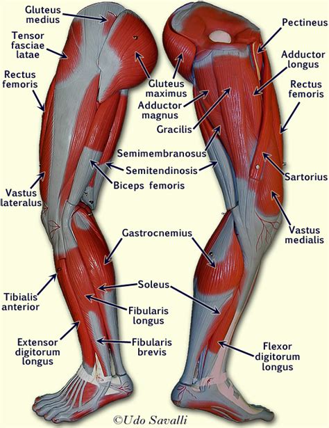
Table of Contents
- Correctly Label The Muscles Of The Leg
- Table of Contents
- Correctly Label the Muscles of the Leg: A Comprehensive Guide
- I. The Muscles of the Thigh
- A. Anterior Compartment (Extensors)
- B. Medial Compartment (Adductors)
- C. Posterior Compartment (Flexors and Rotators)
- II. The Muscles of the Leg
- A. Anterior Compartment (Dorsiflexors)
- B. Lateral Compartment (Evertors)
- C. Posterior Compartment (Plantarflexors)
- III. The Muscles of the Foot
- A. Dorsal Muscles (Top of the Foot)
- B. Plantar Muscles (Sole of the Foot)
- IV. Clinical Significance and Applications
- V. Conclusion
- Latest Posts
- Latest Posts
- Related Post
Correctly Label the Muscles of the Leg: A Comprehensive Guide
Understanding the intricate network of muscles in the leg is crucial for athletes, physical therapists, medical professionals, and anyone interested in human anatomy. This comprehensive guide will delve into the leg's muscular system, providing detailed descriptions and visual aids to help you correctly label each muscle. We'll explore the muscles of the thigh, leg, and foot, categorizing them by function and location for a clear and organized understanding. This guide emphasizes both anatomical precision and practical application, making it useful for students and professionals alike.
I. The Muscles of the Thigh
The thigh muscles are broadly classified into three compartments: anterior (front), medial (inner), and posterior (back).
A. Anterior Compartment (Extensors)
The anterior compartment muscles primarily extend the leg at the knee and flex the hip. They are innervated by the femoral nerve.
-
Quadriceps Femoris: This powerful group consists of four muscles:
- Rectus Femoris: Originates from the anterior inferior iliac spine and superior acetabulum; inserts into the tibial tuberosity via the patellar tendon. It's the only quadriceps muscle that crosses both the hip and knee joints, allowing for both hip flexion and knee extension. Key Action: Hip flexion, knee extension.
- Vastus Lateralis: The largest of the quadriceps; originates from the greater trochanter, intertrochanteric line, and linea aspera of the femur; inserts into the tibial tuberosity via the patellar tendon. Key Action: Knee extension.
- Vastus Medialis: Located on the medial aspect of the thigh; originates from the intertrochanteric line and medial lip of the linea aspera; inserts into the tibial tuberosity via the patellar tendon. Key Action: Knee extension.
- Vastus Intermedius: Deep to the rectus femoris; originates from the anterior and lateral surfaces of the femur; inserts into the tibial tuberosity via the patellar tendon. Key Action: Knee extension.
-
Sartorius: The longest muscle in the body; originates from the anterior superior iliac spine; inserts into the medial surface of the tibia. It acts as a weak flexor of the hip and knee, and also assists in abduction and lateral rotation of the thigh. Key Action: Hip flexion, abduction, lateral rotation; knee flexion.
B. Medial Compartment (Adductors)
The medial compartment muscles primarily adduct the thigh (bring it towards the midline). They are innervated by the obturator nerve (with some exceptions).
- Adductor Longus: Originates from the pubic symphysis; inserts into the linea aspera of the femur. Key Action: Hip adduction, flexion, medial rotation.
- Adductor Brevis: Located deep to the adductor longus; originates from the pubis; inserts into the linea aspera of the femur. Key Action: Hip adduction, flexion, medial rotation.
- Adductor Magnus: The largest of the adductors; has two heads – an adductor part and a hamstring part. Originates from the pubis and ischial tuberosity; inserts into the linea aspera and adductor tubercle of the femur. Key Action: Hip adduction, extension (hamstring part), medial rotation.
- Gracilis: A long, slender muscle; originates from the pubic symphysis; inserts into the medial surface of the tibia. Key Action: Hip adduction; knee flexion.
- Pectineus: Originates from the pectineal line of the pubis; inserts into the pectineal line of the femur. Key Action: Hip flexion, adduction.
C. Posterior Compartment (Flexors and Rotators)
The posterior compartment muscles primarily flex the leg at the knee and extend the hip. They are innervated by the sciatic nerve (which branches into the tibial and common peroneal nerves).
- Hamstrings: This group consists of three muscles:
- Biceps Femoris: Has two heads – a long head originating from the ischial tuberosity and a short head originating from the linea aspera; inserts into the head of the fibula and lateral condyle of the tibia. Key Action: Hip extension, knee flexion, lateral rotation of the leg.
- Semitendinosus: Originates from the ischial tuberosity; inserts into the medial surface of the tibia. Key Action: Hip extension, knee flexion, medial rotation of the leg.
- Semimembranosus: Originates from the ischial tuberosity; inserts into the medial condyle of the tibia. Key Action: Hip extension, knee flexion, medial rotation of the leg.
II. The Muscles of the Leg
The leg muscles are divided into three compartments: anterior (front), lateral (outer), and posterior (back).
A. Anterior Compartment (Dorsiflexors)
The anterior compartment muscles primarily dorsiflex the foot (bring the toes towards the shin). They are innervated by the deep peroneal nerve.
- Tibialis Anterior: Originates from the lateral condyle and upper two-thirds of the tibia; inserts into the medial cuneiform and first metatarsal bones. Key Action: Dorsiflexion, inversion of the foot.
- Extensor Hallucis Longus: Originates from the middle portion of the fibula; inserts into the distal phalanx of the great toe. Key Action: Extension of the great toe, dorsiflexion of the foot.
- Extensor Digitorum Longus: Originates from the lateral condyle of the tibia and the anterior surface of the fibula; inserts into the distal phalanges of the second to fifth toes. Key Action: Extension of the toes, dorsiflexion of the foot.
- Peroneus Tertius: Often considered part of the extensor digitorum longus; inserts into the base of the fifth metatarsal. Key Action: Dorsiflexion, eversion of the foot.
B. Lateral Compartment (Evertors)
The lateral compartment muscles primarily evert the foot (turn the sole away from the midline). They are innervated by the superficial peroneal nerve.
- Peroneus Longus: Originates from the head and upper two-thirds of the fibula; inserts into the medial cuneiform and first metatarsal bones. Key Action: Eversion, plantarflexion of the foot.
- Peroneus Brevis: Originates from the distal third of the fibula; inserts into the base of the fifth metatarsal. Key Action: Eversion, plantarflexion of the foot.
C. Posterior Compartment (Plantarflexors)
The posterior compartment muscles primarily plantarflex the foot (point the toes downwards). They are innervated by the tibial nerve. This compartment is further subdivided into superficial and deep layers.
-
Superficial Layer:
- Gastrocnemius: The superficial muscle of the calf; has two heads originating from the medial and lateral condyles of the femur; inserts into the calcaneus via the Achilles tendon. Key Action: Plantarflexion of the foot, knee flexion.
- Soleus: A deep muscle of the calf; originates from the head and upper third of the fibula and the medial border of the tibia; inserts into the calcaneus via the Achilles tendon. Key Action: Plantarflexion of the foot.
- Plantaris: A small muscle; originates from the lateral supracondylar line of the femur; inserts into the calcaneus via the Achilles tendon. Its function is minimal. Key Action: Weak plantarflexion of the foot, knee flexion.
-
Deep Layer:
- Popliteus: Originates from the lateral condyle of the femur; inserts into the posterior surface of the tibia. Key Action: Knee flexion, medial rotation of the tibia.
- Tibialis Posterior: Originates from the posterior surface of the tibia and fibula; inserts into the navicular, cuneiforms, and cuboid bones. Key Action: Plantarflexion, inversion of the foot.
- Flexor Hallucis Longus: Originates from the posterior surface of the fibula; inserts into the distal phalanx of the great toe. Key Action: Flexion of the great toe, plantarflexion of the foot.
- Flexor Digitorum Longus: Originates from the posterior surface of the tibia; inserts into the distal phalanges of the second to fifth toes. Key Action: Flexion of the toes, plantarflexion of the foot.
III. The Muscles of the Foot
The muscles of the foot are largely intrinsic (located within the foot itself) and are responsible for fine motor control of the toes and arch support. They are innervated by branches of the tibial and deep peroneal nerves. Due to their complexity and numerous small muscles, a detailed description of each is beyond the scope of this introductory guide, but understanding their general functions is important.
A. Dorsal Muscles (Top of the Foot)
These muscles are relatively few compared to the plantar muscles. They are mainly involved in extending the toes.
B. Plantar Muscles (Sole of the Foot)
These muscles are divided into several layers and are responsible for a variety of movements, including flexion of the toes, arch support, and adjustments to gait.
IV. Clinical Significance and Applications
Accurate knowledge of leg muscle anatomy is paramount in various fields:
- Physical Therapy: Therapists use this knowledge to diagnose and treat musculoskeletal injuries, develop targeted exercises, and assess functional limitations.
- Sports Medicine: Understanding muscle function is crucial for injury prevention, rehabilitation, and optimizing athletic performance.
- Orthopedics: Surgeons rely on a deep understanding of muscle attachments and relationships during surgical procedures.
- Neurology: Neurological conditions often manifest as muscle weakness or paralysis, requiring precise anatomical knowledge for diagnosis and management.
V. Conclusion
Mastering the correct labeling of leg muscles requires diligent study and practice. This guide provides a strong foundation for understanding the complex anatomy of the leg. Through consistent review and practical application, you can enhance your comprehension and improve your ability to accurately identify and label each muscle. Remember to utilize anatomical models, diagrams, and interactive resources to aid in your learning journey. The more you engage with this material, the more your knowledge will solidify, paving the way for a deeper understanding of human movement and function. This detailed exploration serves not only as an educational resource but as a tool for professionals to refine their skills and enhance their clinical practice. The ability to accurately identify and understand the functions of these muscles is fundamental to effective diagnosis, treatment, and overall patient care.
Latest Posts
Latest Posts
-
Ribosomal Subunits Are Manufactured By The
Mar 30, 2025
-
Accounts Receivable Are Normally Reported At The
Mar 30, 2025
-
Unlike Eutherians Both Monotremes And Marsupials
Mar 30, 2025
-
Draw The Enantiomer Of The Molecule Shown Below
Mar 30, 2025
-
When A Union Bargains Successfully With Employers In That Industry
Mar 30, 2025
Related Post
Thank you for visiting our website which covers about Correctly Label The Muscles Of The Leg . We hope the information provided has been useful to you. Feel free to contact us if you have any questions or need further assistance. See you next time and don't miss to bookmark.
