Correctly Label The Intrinsic Muscles Of The Hand
Holbox
Mar 26, 2025 · 5 min read
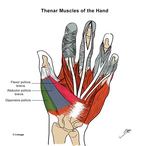
Table of Contents
- Correctly Label The Intrinsic Muscles Of The Hand
- Table of Contents
- Correctly Labeling the Intrinsic Muscles of the Hand: A Comprehensive Guide
- Understanding the Organization of Intrinsic Hand Muscles
- 1. Thenar Muscles (Thumb Muscles):
- 2. Hypothenar Muscles (Little Finger Muscles):
- 3. Midpalmar Muscles:
- Mnemonic Devices for Easier Memorization
- Clinical Correlations and Importance of Accurate Labeling
- Advanced Considerations: Variations and Anatomic Details
- Further Learning and Resources
- Conclusion
- Latest Posts
- Latest Posts
- Related Post
Correctly Labeling the Intrinsic Muscles of the Hand: A Comprehensive Guide
The intrinsic muscles of the hand are a complex group of muscles residing entirely within the hand, responsible for fine motor control, dexterity, and the intricate movements that make our hands so versatile. Correctly identifying and labeling these muscles is crucial for healthcare professionals, anatomy students, and anyone interested in understanding the intricacies of hand function. This comprehensive guide will break down each muscle, providing clear descriptions, origins, insertions, actions, and innervations. We'll also explore common clinical correlations and mnemonic devices to aid in memorization.
Understanding the Organization of Intrinsic Hand Muscles
Before diving into individual muscles, it's helpful to understand their organization. The intrinsic hand muscles are broadly classified into three groups based on their location and function:
1. Thenar Muscles (Thumb Muscles):
These muscles are located in the thenar eminence, the fleshy mound at the base of the thumb. They are responsible for thumb opposition, flexion, abduction, and adduction. They include:
-
Abductor pollicis brevis: Origin: Scaphoid and trapezium. Insertion: Radial side of proximal phalanx of thumb. Action: Abducts the thumb. Innervation: Median nerve.
-
Flexor pollicis brevis: Origin: Trapezium and flexor retinaculum. Insertion: Radial side of proximal phalanx of thumb. Action: Flexes the thumb. Innervation: Median nerve. Note: It has a superficial and deep head, often differentiated in detailed anatomical studies.
-
Opponens pollicis: Origin: Trapezium. Insertion: Radial side of first metacarpal. Action: Opposes the thumb (brings it across the palm towards the little finger). Innervation: Median nerve.
-
Adductor pollicis: Origin: Carpal bones (capitate and bases of second and third metacarpals). Insertion: Ulnar side of proximal phalanx of thumb. Action: Adducts the thumb. Innervation: Deep branch of ulnar nerve. (This is unique as it's innervated by the ulnar nerve, unlike the other thenar muscles).
2. Hypothenar Muscles (Little Finger Muscles):
These muscles reside in the hypothenar eminence, the fleshy mound at the base of the little finger. They control the little finger's movement. They include:
-
Abductor digiti minimi: Origin: Pisiform bone and flexor retinaculum. Insertion: Ulnar side of proximal phalanx of little finger. Action: Abducts the little finger. Innervation: Ulnar nerve.
-
Flexor digiti minimi brevis: Origin: Hamate and flexor retinaculum. Insertion: Ulnar side of proximal phalanx of little finger. Action: Flexes the little finger. Innervation: Ulnar nerve.
-
Opponens digiti minimi: Origin: Hamate and flexor retinaculum. Insertion: Ulnar side of fifth metacarpal. Action: Opposes the little finger (brings it towards the thumb). Innervation: Ulnar nerve.
3. Midpalmar Muscles:
This group includes the lumbricals, interossei, and the palmar and dorsal interossei muscles. These muscles are responsible for finger flexion, extension, abduction, and adduction. They are crucial for complex hand movements.
-
Lumbricals (4 muscles): Origin: Tendons of flexor digitorum profundus. Insertion: Radial side of extensor expansion of the index, middle, ring, and little fingers. Action: Flexes the metacarpophalangeal joints and extends the interphalangeal joints of fingers 2-5. Innervation: Lumbricals 1 and 2 are innervated by the median nerve, while lumbricals 3 and 4 are innervated by the ulnar nerve.
-
Palmar Interossei (3 muscles): Origin: Sides of metacarpals (2,4,5). Insertion: Proximal phalanges and extensor expansions of fingers 2, 4, and 5. Action: Adduct fingers 2, 4, and 5 towards the middle finger. Innervation: Deep branch of ulnar nerve.
-
Dorsal Interossei (4 muscles): Origin: Between metacarpals. Insertion: Proximal phalanges and extensor expansions of fingers 2, 3, and 4. Action: Abduct fingers 2, 3, and 4 away from the middle finger. Innervation: Deep branch of ulnar nerve.
Mnemonic Devices for Easier Memorization
Learning the intrinsic muscles of the hand can be challenging due to their number and subtle anatomical differences. Mnemonic devices can significantly aid in memorization:
-
Thenar Muscles: Think "All Fingers Oppose Advanced Playing" - Abductor pollicis brevis, Flexor pollicis brevis, Opponens pollicis, Adductor pollicis.
-
Hypothenar Muscles: "All Fingers Oppose Hand" - Abductor digiti minimi, Flexor digiti minimi brevis, Opponens digiti minimi. (Adding the "H" for Hypothenar).
-
Interossei: Remember that the palmar interossei adduct (move towards the middle finger) while the dorsal interossei abduct (move away from the middle finger).
Clinical Correlations and Importance of Accurate Labeling
Accurate labeling of the intrinsic hand muscles is paramount in various clinical settings:
-
Diagnosis of Nerve Injuries: Knowing the innervation of each muscle allows clinicians to pinpoint the location and severity of nerve damage based on muscle weakness or paralysis. For example, weakness in the thenar muscles suggests median nerve involvement, while weakness in the hypothenar and interossei muscles points towards ulnar nerve injury.
-
Surgical Procedures: Surgeons rely on precise anatomical knowledge to perform hand surgeries successfully, minimizing complications and maximizing functional recovery. Accurate labeling is essential for tendon repairs, nerve grafts, and other procedures involving the intrinsic hand muscles.
-
Rehabilitation: Physical and occupational therapists utilize their understanding of these muscles to design effective rehabilitation programs for patients with hand injuries or conditions. Proper identification helps in targeted exercises and functional assessments.
-
Assessment of Hand Function: Clinicians assess hand function by evaluating the strength and range of motion of individual muscles, providing a detailed picture of hand health and dysfunction.
Advanced Considerations: Variations and Anatomic Details
It’s important to remember that anatomical variations exist. The size and precise attachments of these muscles can differ between individuals. Furthermore, some muscles might have additional slips or be partially fused with neighboring muscles. Detailed anatomical dissections and imaging studies are crucial for understanding these individual differences. Precise clinical assessment relies on comprehensive knowledge of potential variations.
Further Learning and Resources
This guide provides a foundation for understanding the intrinsic muscles of the hand. For deeper exploration, consult detailed anatomical textbooks, atlases, and online resources. Engaging with three-dimensional anatomical models and interactive simulations can greatly enhance learning and retention. Consider supplementing your study with practical applications, such as observing anatomical dissections (under appropriate supervision), participating in hands-on clinical experiences, and engaging with case studies that highlight clinical correlations.
Conclusion
The intrinsic muscles of the hand are a fascinating and complex system that enables the remarkable dexterity and fine motor control characteristic of human hands. Accurate labeling and understanding of these muscles are vital for healthcare professionals, researchers, and anyone interested in the human anatomy. By utilizing the information and mnemonic devices in this guide and supplementing with additional learning resources, one can develop a strong understanding of this intricate muscular system. Remember, the key to mastering this topic lies in consistent study, practical application, and a genuine appreciation for the complexities and elegance of human anatomy.
Latest Posts
Latest Posts
-
Which Helps Enable An Oligopoly To Form Within A Market
Mar 29, 2025
-
Lagle Corporation Has Provided The Following Information
Mar 29, 2025
-
The Clear Zone Around An Antibiotic Disk
Mar 29, 2025
-
Experiment 25 Ph Measurements Buffers And Their Properties
Mar 29, 2025
-
What Should Managers Do When It Comes To Stress Reduction
Mar 29, 2025
Related Post
Thank you for visiting our website which covers about Correctly Label The Intrinsic Muscles Of The Hand . We hope the information provided has been useful to you. Feel free to contact us if you have any questions or need further assistance. See you next time and don't miss to bookmark.
