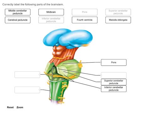
Correctly Label The Following Parts Of The Brainstem
Holbox
Mar 31, 2025 · 6 min read

Table of Contents
- Correctly Label The Following Parts Of The Brainstem
- Table of Contents
- Correctly Label the Following Parts of the Brainstem: A Comprehensive Guide
- Major Divisions of the Brainstem
- 1. The Midbrain (Mesencephalon)
- 2. The Pons (Metencephalon)
- 3. The Medulla Oblongata (Myelencephalon)
- Labeling the Brainstem: A Practical Approach
- Beyond Basic Labeling: Understanding the Functional Interconnections
- Conclusion
- Latest Posts
- Latest Posts
- Related Post
Correctly Label the Following Parts of the Brainstem: A Comprehensive Guide
The brainstem, a crucial structure connecting the cerebrum and cerebellum to the spinal cord, is a complex network responsible for essential life-sustaining functions. Understanding its intricate anatomy is vital for anyone studying neuroscience, medicine, or related fields. This comprehensive guide will delve deep into the brainstem's components, providing a detailed explanation of each part and how to correctly label them. We'll go beyond simple identification, exploring the function and significance of each region.
Major Divisions of the Brainstem
The brainstem is conventionally divided into three main parts: the midbrain, the pons, and the medulla oblongata. Each section possesses unique anatomical features and distinct functional roles, working together seamlessly to maintain homeostasis and regulate vital bodily processes.
1. The Midbrain (Mesencephalon)
The midbrain, the most superior part of the brainstem, is relatively small but packed with critical structures. Key components include:
-
Superior Colliculi: These paired, rounded structures are primarily involved in visual processing, particularly in orienting the eyes and head towards visual stimuli. They act as a relay station for visual reflexes and play a significant role in visually guided movements. Think of them as the brain's "visual router."
-
Inferior Colliculi: Located just below the superior colliculi, these structures are primarily involved in auditory processing. They receive auditory input from the cochlea and relay it to other brain regions involved in hearing and sound localization. They're crucial for determining the direction and source of sounds.
-
Substantia Nigra: This darkly pigmented structure plays a vital role in movement control. Its neurons produce dopamine, a neurotransmitter essential for smooth, coordinated muscle movement. Degeneration of dopaminergic neurons in the substantia nigra is a hallmark of Parkinson's disease.
-
Red Nucleus: This reddish-colored nucleus (due to its rich blood supply and iron content) is involved in motor coordination. It's particularly crucial for the fine motor control of the limbs. It interacts closely with the cerebellum and other motor centers.
-
Cerebral Peduncles: These large fiber tracts on the ventral surface of the midbrain connect the cerebrum to the pons and cerebellum. They are crucial for conveying motor commands from the cortex to the lower motor neurons in the brainstem and spinal cord.
Clinical Significance: Damage to the midbrain can result in a range of neurological deficits, including impaired vision, hearing loss, movement disorders (like Parkinsonism), and oculomotor problems (difficulty controlling eye movements).
2. The Pons (Metencephalon)
The pons, situated below the midbrain, is a bulging structure that acts as a bridge between different parts of the brain. Its key features include:
-
Pontine Nuclei: These large clusters of neurons receive input from the cerebral cortex and relay this information to the cerebellum via the transverse pontine fibers. This pathway is crucial for coordinating voluntary movements. Think of the pontine nuclei as the "message center" between the cerebrum and cerebellum.
-
Cranial Nerve Nuclei: Several cranial nerves (V, VI, VII, and VIII) originate within the pons, controlling functions such as facial expression, eye movement, hearing, and balance. Damage to these nuclei can cause various cranial nerve palsies, resulting in facial weakness, hearing loss, or balance problems.
-
Respiratory Centers: The pons plays a crucial role in regulating respiration. It contains nuclei that modify the respiratory rhythm generated by the medulla oblongata, helping to fine-tune breathing patterns according to the body's needs.
Clinical Significance: Damage to the pons can lead to a range of symptoms, including respiratory dysfunction, problems with balance and coordination, loss of facial sensation, and various cranial nerve palsies. Locked-in syndrome, a rare condition where patients are conscious but unable to move or communicate except through eye movements, can result from pontine damage.
3. The Medulla Oblongata (Myelencephalon)
The medulla oblongata, the most caudal part of the brainstem, is continuous with the spinal cord. This vital region controls many essential autonomic functions:
-
Pyramids: These prominent bulges on the ventral surface of the medulla contain the corticospinal tracts, which carry motor commands from the cerebral cortex to the spinal cord. The decussation of the pyramids, where the motor fibers cross over to the opposite side, is a significant anatomical landmark.
-
Olives: These oval-shaped structures located on the lateral surface of the medulla are involved in motor learning and coordination. They receive sensory input from the spinal cord and cerebellum and project to the cerebellum, contributing to motor control.
-
Cranial Nerve Nuclei: Several cranial nerves (IX, X, XI, and XII) originate in the medulla, controlling functions such as swallowing, speech, head and neck movement, and visceral functions (like heart rate and blood pressure).
-
Vital Centers: The medulla contains clusters of neurons that regulate vital autonomic functions, including:
- Cardiovascular Center: Controls heart rate and blood pressure.
- Respiratory Center: Generates the basic rhythm of breathing (in coordination with the pons).
- Vasomotor Center: Regulates blood vessel diameter, influencing blood pressure.
Clinical Significance: Damage to the medulla oblongata is often life-threatening, potentially causing respiratory arrest, cardiovascular collapse, and disruption of other vital functions. Even minor damage can lead to significant neurological deficits.
Labeling the Brainstem: A Practical Approach
Accurately labeling the brainstem requires careful observation and a thorough understanding of its anatomy. Here’s a step-by-step approach:
-
Start with the major divisions: Begin by clearly identifying the midbrain, pons, and medulla oblongata. Look for the clear anatomical boundaries between these regions.
-
Identify key landmarks: Focus on readily identifiable structures like the pyramids, olives, superior and inferior colliculi, and cerebral peduncles. These serve as excellent reference points.
-
Use cross-sectional diagrams: Studying cross-sections of the brainstem can be highly beneficial. These diagrams often show the internal structures and their spatial relationships more clearly.
-
Consult anatomical atlases: Utilize reliable anatomical atlases or textbooks to verify your labeling and gain a deeper understanding of the structures' relationships.
-
Practice, practice, practice: Repeatedly labeling diagrams and studying the brainstem's anatomy will greatly improve your accuracy and confidence.
Beyond Basic Labeling: Understanding the Functional Interconnections
Correctly labeling the brainstem's components is just the first step. A deeper understanding requires grasping the intricate functional interconnections between its various parts. For example:
-
The relationship between the midbrain and cerebellum: The midbrain's red nucleus interacts closely with the cerebellum in coordinating movement.
-
The role of the pons in coordinating movement: The pontine nuclei act as a crucial relay station between the cerebrum and cerebellum, essential for smooth, coordinated motor control.
-
The medulla's control over autonomic functions: The vital centers in the medulla are responsible for regulating essential life-sustaining processes, making it a critical area for survival.
Conclusion
The brainstem is a vital structure with complex anatomy and critical functions. Correctly labeling its components is essential for comprehending its role in maintaining homeostasis and regulating vital bodily processes. This comprehensive guide has provided a detailed exploration of the brainstem's major regions, their functions, and clinical significance. By combining careful study, utilizing anatomical resources, and understanding functional interconnections, one can achieve mastery in labeling and understanding this crucial part of the central nervous system. Remember, consistent practice and a curious approach will solidify your knowledge and significantly enhance your understanding of neuroanatomy. Continue exploring, and your comprehension of this intricate structure will deepen!
Latest Posts
Latest Posts
-
Draw Cis 1 Ethyl 2 Isopropylcyclohexane In Its Lowest Energy Conformation
Apr 03, 2025
-
Which Of The Following Is A Limitation Of Activity Based Costing
Apr 03, 2025
-
Which Code Example Is An Expression
Apr 03, 2025
-
Good Team Goals Are All Of The Following Except
Apr 03, 2025
-
During The Year Trc Corporation Has The Following Inventory Transactions
Apr 03, 2025
Related Post
Thank you for visiting our website which covers about Correctly Label The Following Parts Of The Brainstem . We hope the information provided has been useful to you. Feel free to contact us if you have any questions or need further assistance. See you next time and don't miss to bookmark.
