Correctly Label The Following External Anatomy Of The Anterior Heart.
Holbox
Mar 30, 2025 · 5 min read
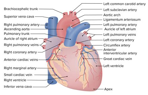
Table of Contents
- Correctly Label The Following External Anatomy Of The Anterior Heart.
- Table of Contents
- Correctly Labeling the External Anatomy of the Anterior Heart
- Locating the Heart and Establishing Orientation
- Key External Structures of the Anterior Heart
- 1. Right Atrium (RA):
- 2. Right Ventricle (RV):
- 3. Left Ventricle (LV):
- 4. Left Atrium (LA):
- 5. Auricles (Atrial Appendages):
- 6. Sulci (Grooves):
- Understanding the Coronary Vessels (Visible in Sulci)
- Clinical Significance of Correctly Identifying Anterior Heart Anatomy
- Tips for Effective Learning and Labeling
- Latest Posts
- Latest Posts
- Related Post
Correctly Labeling the External Anatomy of the Anterior Heart
The human heart, a remarkable organ, tirelessly pumps blood throughout our bodies. Understanding its anatomy, particularly the external features visible on its anterior (front) surface, is crucial for anyone studying anatomy, physiology, or related medical fields. This comprehensive guide will walk you through the process of correctly labeling the external anatomy of the anterior heart, providing detailed descriptions and high-yield information for effective learning and recall.
Locating the Heart and Establishing Orientation
Before we begin labeling the specific structures, it's essential to understand the heart's location and orientation within the thoracic cavity. The heart lies within the mediastinum, a central compartment of the chest, slightly angled to the left. Its apex (pointed end) points inferiorly and slightly to the left, while its base (broader end) lies superiorly and posteriorly. This orientation is critical when visualizing and labeling the various structures.
Remember that we're focusing on the anterior surface—the aspect facing forward when the body is in anatomical position.
Key External Structures of the Anterior Heart
Now, let's dive into the specific structures visible on the anterior surface of the heart:
1. Right Atrium (RA):
- Location: Forms a significant portion of the heart's right border. A small portion might be visible anteriorly, depending on the angle of view.
- Function: Receives deoxygenated blood returning from the body through the superior and inferior vena cava.
- Identifying Features: Often less prominent on the anterior surface compared to other chambers. You may see a faint sulcus (groove) separating it from the right ventricle.
2. Right Ventricle (RV):
- Location: Dominates the anterior surface of the heart. Its muscular wall is relatively thin compared to the left ventricle.
- Function: Pumps deoxygenated blood to the lungs via the pulmonary artery.
- Identifying Features: Forms the most prominent bulge on the anterior surface. You'll likely notice the anterior interventricular sulcus running diagonally across its surface. This groove houses the anterior interventricular artery (a branch of the left coronary artery).
3. Left Ventricle (LV):
- Location: A significant portion lies posteriorly, but a smaller part is visible on the anterior surface, usually at the leftmost part of the heart.
- Function: Pumps oxygenated blood to the entire body through the aorta.
- Identifying Features: Though less visible anteriorly compared to the RV, the LV's thicker muscular wall contributes to the overall shape and robustness of the heart's apex. The anterior interventricular sulcus also marks its border with the RV.
4. Left Atrium (LA):
- Location: Primarily located on the heart's posterior surface and largely hidden from view on the anterior surface. A small portion might be visible near the base of the heart, particularly if the surrounding structures are carefully removed or visualized in an image.
- Function: Receives oxygenated blood from the lungs via the pulmonary veins.
- Identifying Features: Difficult to distinctly identify on the anterior aspect. Its presence is more inferred based on the overall heart shape and neighboring structures.
5. Auricles (Atrial Appendages):
- Location: Small, ear-like extensions protruding from the atria. The right auricle is more visible on the anterior surface than the left.
- Function: Increase the atrial volume slightly, aiding in blood collection.
- Identifying Features: Their shape is easily distinguishable. Look for small pouch-like structures extending from the atria.
6. Sulci (Grooves):
- Location: These are external grooves that mark the boundaries between the heart chambers.
- Function: Contain coronary arteries and veins.
- Identifying Features: The anterior interventricular sulcus is the most prominent, running diagonally down the anterior surface. A less distinct atrioventricular sulcus encircles the heart, separating the atria from the ventricles. These sulci are crucial landmarks for understanding the heart's internal structure.
Understanding the Coronary Vessels (Visible in Sulci)
The coronary arteries and veins are essential for supplying the heart muscle itself with oxygen and nutrients. These vessels are typically found within the sulci, and while not strictly part of the heart's chambers, their presence on the anterior surface is crucial for a complete anatomical understanding.
- Anterior Interventricular Artery (a branch of the left coronary artery): Located within the anterior interventricular sulcus. Supplies blood to the anterior portion of both ventricles.
- Circumflex Artery (a branch of the left coronary artery): Often partially visible in the atrioventricular sulcus. Supplies blood to the left atrium and parts of the left ventricle.
- Right Coronary Artery: May be visible in the atrioventricular sulcus. Supplies blood to the right atrium, right ventricle, and parts of the left ventricle and posterior heart.
- Great Cardiac Vein: Often visible in the anterior interventricular sulcus. Collects blood from the anterior heart.
- Anterior Cardiac Veins: Several smaller veins that drain into the great cardiac vein.
Clinical Significance of Correctly Identifying Anterior Heart Anatomy
Accurate identification of the anterior heart's external anatomy is crucial in various clinical settings:
- Cardiac Catheterization: Understanding the location of coronary arteries and veins is essential for performing cardiac catheterization procedures, such as angioplasty and stenting.
- Cardiac Surgery: Surgical procedures requiring access to the heart depend on precise anatomical knowledge to minimize complications.
- Electrocardiography (ECG): Understanding heart anatomy helps in interpreting ECG results and diagnosing cardiac conditions.
- Cardiac Imaging: Interpreting images such as echocardiograms and cardiac CT scans necessitates a strong foundation in heart anatomy.
Tips for Effective Learning and Labeling
Mastering the external anatomy of the anterior heart requires consistent effort and a multi-faceted approach:
- Use Anatomical Models: Physical models allow for hands-on learning and a three-dimensional understanding of the heart's structure.
- Refer to High-Quality Images: Use anatomical atlases, textbooks, or online resources with detailed illustrations.
- Labeling Practice: Repeatedly label diagrams and images of the anterior heart. This reinforces your knowledge and helps with memorization.
- Clinical Correlation: Connecting anatomical knowledge to clinical scenarios enhances understanding and retention.
- Peer Learning: Discuss and quiz each other with classmates or colleagues to improve comprehension and identify areas needing further study.
- Utilize Flashcards: Create flashcards with images and corresponding labels to aid in memorization. Include both the name and function of each structure for comprehensive learning.
- Employ Mnemonics: Develop mnemonics or memory aids to assist in remembering the locations and functions of various structures.
By following these strategies, you can effectively learn and confidently label the external anatomy of the anterior heart. Remember that consistent effort and a multi-sensory approach are key to mastering this essential anatomical concept. Good luck!
Latest Posts
Latest Posts
-
Drag Each Label To The Type Of Gland It Describes
Apr 02, 2025
-
Name The Membranous Encasement Surrounding The Brain
Apr 02, 2025
-
Select The Statements About The K T Boundary That Are True
Apr 02, 2025
-
All Of The Following Are Examples Of Product Costs Except
Apr 02, 2025
-
Which Of These Is Unique To Flowering Plants
Apr 02, 2025
Related Post
Thank you for visiting our website which covers about Correctly Label The Following External Anatomy Of The Anterior Heart. . We hope the information provided has been useful to you. Feel free to contact us if you have any questions or need further assistance. See you next time and don't miss to bookmark.
