Correctly Label The Following Anatomical Features Of The Tibiofemoral Joint
Holbox
Mar 21, 2025 · 6 min read
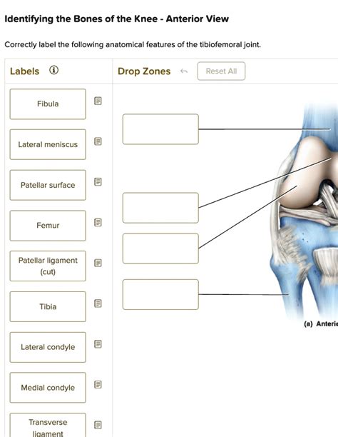
Table of Contents
Correctly Labeling the Anatomical Features of the Tibiofemoral Joint
The tibiofemoral joint, commonly known as the knee joint, is one of the largest and most complex joints in the human body. Its intricate structure allows for a wide range of motion, crucial for activities like walking, running, jumping, and squatting. Understanding its anatomy is essential for anyone studying anatomy, physiotherapy, orthopedics, or related fields. This comprehensive guide will walk you through the correct labeling of the key anatomical features of the tibiofemoral joint, providing detailed descriptions and clarifying common points of confusion.
Bones of the Tibiofemoral Joint
The tibiofemoral joint is formed by the articulation of three bones:
1. Femur (Thigh Bone)
The distal end of the femur, the thigh bone, plays a crucial role in the knee joint. Key features to identify include:
- Medial Condyle: The larger, medial (inner) condyle of the femur articulates with the medial tibial condyle. Its smooth articular surface contributes to the stability and movement of the knee.
- Lateral Condyle: The slightly smaller, lateral (outer) condyle of the femur articulates with the lateral tibial condyle. Similar to the medial condyle, its articular surface is crucial for joint function.
- Intercondylar Notch (Intercondylar Fossa): Located between the medial and lateral condyles, this notch houses the crucial cruciate ligaments, providing essential stability to the knee.
- Epicondyles (Medial and Lateral): These bony prominences on the sides of the condyles serve as attachment points for several important muscles and ligaments of the knee, contributing to its overall stability and movement.
2. Tibia (Shin Bone)
The proximal end of the tibia, the shin bone, is the main weight-bearing bone of the lower leg and a key component of the tibiofemoral joint. Key features to identify are:
- Medial Condyle: This broad, flat articular surface on the medial side of the tibia articulates with the medial condyle of the femur.
- Lateral Condyle: This similarly shaped articular surface on the lateral side of the tibia articulates with the lateral condyle of the femur.
- Intercondylar Eminence: This raised area between the medial and lateral condyles serves as the attachment point for the anterior and posterior cruciate ligaments. It's crucial for the stability of the knee joint.
- Tibial Tuberosity: Located on the anterior surface of the tibia, inferior to the intercondylar eminence, this roughened area serves as the attachment point for the patellar ligament. This ligament connects the patella (kneecap) to the tibia.
3. Patella (Kneecap)
The patella, a sesamoid bone, is embedded within the quadriceps tendon and articulates with the patellar surface of the femur. Correct labeling requires understanding its articulation and relationship to the other bones:
- Articular Surface: The posterior surface of the patella is covered with articular cartilage and articulates with the patellar surface of the femur, providing smooth movement during knee flexion and extension.
- Apex: The pointed distal end of the patella.
- Base: The broader proximal end of the patella.
Ligaments of the Tibiofemoral Joint
The stability of the tibiofemoral joint heavily relies on a complex network of ligaments:
1. Cruciate Ligaments (Intracapsular)
These ligaments are located within the joint capsule and cross each other, forming an "X" shape. They're crucial for controlling anterior and posterior movement of the tibia relative to the femur.
- Anterior Cruciate Ligament (ACL): Prevents anterior translation of the tibia on the femur. It attaches to the anterior intercondylar area of the tibia and the lateral aspect of the medial femoral condyle.
- Posterior Cruciate Ligament (PCL): Prevents posterior translation of the tibia on the femur. It attaches to the posterior intercondylar area of the tibia and the medial aspect of the lateral femoral condyle.
2. Collateral Ligaments (Extracapsular)
These ligaments are located outside the joint capsule and provide medial and lateral stability.
- Medial Collateral Ligament (MCL): A broad, flat ligament extending from the medial epicondyle of the femur to the medial tibial condyle. It prevents excessive valgus stress (medial movement) on the knee.
- Lateral Collateral Ligament (LCL): A cord-like ligament extending from the lateral epicondyle of the femur to the head of the fibula. It prevents excessive varus stress (lateral movement) on the knee.
Menisci of the Tibiofemoral Joint
The menisci are two C-shaped fibrocartilaginous structures located between the tibial and femoral condyles. They act as shock absorbers, distributing weight evenly across the joint and enhancing stability.
- Medial Meniscus: C-shaped, larger and more firmly attached to the tibial capsule. More prone to injury due to its less mobile nature.
- Lateral Meniscus: More circular in shape and more mobile than the medial meniscus.
Articular Cartilage
The articular cartilage covering the articular surfaces of the femur, tibia, and patella is essential for smooth, low-friction movement within the joint. Damage to this cartilage can lead to osteoarthritis.
Bursae of the Tibiofemoral Joint
Several bursae, fluid-filled sacs, are located around the knee joint. They reduce friction between tendons, ligaments, and bones. Correct identification requires understanding their locations and functions. Some of the most significant include the prepatellar bursa, infrapatellar bursa, and suprapatellar bursa.
Muscles Acting on the Tibiofemoral Joint
A vast array of muscles contributes to the movement and stability of the knee joint. Proper labeling necessitates understanding their origin, insertion, and actions. Key muscle groups include:
- Quadriceps Femoris: Rectus femoris, vastus lateralis, vastus medialis, and vastus intermedius—these muscles extend the knee.
- Hamstrings: Biceps femoris, semitendinosus, and semimembranosus—these muscles flex the knee.
- Gastrocnemius: Assists with knee flexion.
- Popliteus: Unlocks the knee and initiates flexion.
Common Errors in Labeling
Several common errors occur when labeling the tibiofemoral joint. These are often due to the joint's complexity and the close proximity of structures.
- Confusing Medial and Lateral: Students often confuse the medial and lateral structures. Remember that "medial" refers to the inner aspect of the limb, while "lateral" refers to the outer aspect.
- Misidentifying Cruciate Ligaments: The anterior and posterior cruciate ligaments are often confused. Pay close attention to their attachment points to avoid this error.
- Incorrect Placement of Menisci: The menisci can be mislabeled or drawn incorrectly. Familiarize yourself with their C-shaped structure and position between the tibial and femoral condyles.
- Overlooking Smaller Structures: Bursae and smaller ligaments are sometimes overlooked. Pay careful attention to detail when labeling.
Importance of Accurate Labeling
Accurately labeling the anatomical features of the tibiofemoral joint is crucial for several reasons:
- Clinical Diagnosis: Accurate anatomical knowledge is essential for clinicians to diagnose and treat knee injuries effectively.
- Surgical Planning: Orthopedic surgeons rely on precise anatomical knowledge for surgical planning and execution.
- Rehabilitation: Physiotherapists use anatomical knowledge to develop effective rehabilitation programs.
- Research: Researchers rely on accurate labeling for consistent and meaningful communication of findings.
- Education: Accurate labeling is critical for students and professionals to build a solid foundation in anatomy and kinesiology.
Conclusion
Mastering the correct labeling of the tibiofemoral joint’s features is a significant step towards a thorough understanding of this complex joint. By focusing on the details, understanding the functional relationships between structures, and diligently practicing labeling techniques, you can achieve a solid grasp of this essential anatomical region. Remember to use high-quality anatomical diagrams and models to aid your learning. Consistent review and application of this knowledge will significantly enhance your comprehension and ability to accurately label all components of the tibiofemoral joint.
Latest Posts
Latest Posts
-
Menlo Company Distributes A Single Product
Mar 22, 2025
-
Which Reaction Sequence Best Accomplishes This Transformation
Mar 22, 2025
-
One Consequence Of Todays High Choice Media System Is
Mar 22, 2025
-
Lab 1 Vertical Structure Of The Atmosphere Answers
Mar 22, 2025
-
Shaping Is A Method Used By Skinner To
Mar 22, 2025
Related Post
Thank you for visiting our website which covers about Correctly Label The Following Anatomical Features Of The Tibiofemoral Joint . We hope the information provided has been useful to you. Feel free to contact us if you have any questions or need further assistance. See you next time and don't miss to bookmark.
