Correctly Label The Following Anatomical Features Of The Spinal Cord
Holbox
Mar 20, 2025 · 6 min read
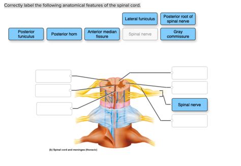
Table of Contents
Correctly Labeling the Anatomical Features of the Spinal Cord: A Comprehensive Guide
The spinal cord, a vital component of the central nervous system, is a complex structure responsible for transmitting information between the brain and the rest of the body. Understanding its intricate anatomy is crucial for anyone studying neuroscience, medicine, or related fields. This comprehensive guide will delve into the key anatomical features of the spinal cord, providing detailed descriptions and assisting in their accurate labeling. We'll explore its external and internal structures, highlighting important landmarks and their functional significance.
External Anatomy of the Spinal Cord: A Macroscopic View
Before diving into the microscopic details, let's first examine the external features visible to the naked eye. The spinal cord, roughly cylindrical in shape, extends from the medulla oblongata of the brainstem to the conus medullaris, typically ending around the L1-L2 vertebral level in adults. Several key external features need careful observation and labeling:
1. Conus Medullaris: The Tapering End
The conus medullaris is the tapered, conical end of the spinal cord. It marks the termination point of the neural tissue itself. Distal to the conus medullaris lies the filum terminale, a slender thread of connective tissue extending from the conus medullaris to the coccyx, providing structural support. Correctly identifying the conus medullaris is essential as it represents the caudal limit of the spinal cord proper.
2. Cauda Equina: The "Horse's Tail"
Inferior to the conus medullaris lies the cauda equina, a collection of nerve roots resembling a horse's tail. These nerve roots extend from the lower lumbar, sacral, and coccygeal segments of the spinal cord, continuing to their respective foramina for exit from the vertebral column. Understanding the cauda equina's arrangement is crucial for procedures such as lumbar puncture (spinal tap), which carefully navigates this region to access cerebrospinal fluid.
3. Cervical and Lumbar Enlargements: Bulges of Neural Tissue
Two noticeable swellings, the cervical enlargement and the lumbar enlargement, are present along the spinal cord. These enlargements correspond to the regions where nerves innervate the upper and lower limbs, respectively. The increased neural tissue in these areas reflects the greater number of neurons required to control the complex motor and sensory functions of the extremities. Precise labeling of these enlargements provides a clear indication of the functional importance of these segments.
4. Spinal Nerves: The Communication Pathways
Emerging from the spinal cord at regular intervals are spinal nerves, each representing a mixed nerve containing both sensory and motor fibers. These nerves exit the vertebral column via the intervertebral foramina. Accurate labeling should include distinguishing between the anterior (ventral) and posterior (dorsal) roots of each spinal nerve. The anterior root contains motor axons, while the posterior root carries sensory axons. The union of these roots forms the spinal nerve.
5. Anterior Median Fissure and Posterior Median Sulcus: External Grooves
The spinal cord exhibits two longitudinal grooves, the anterior median fissure and the posterior median sulcus. These grooves do not completely divide the spinal cord but serve as external landmarks separating the left and right halves. The anterior median fissure is deeper than the posterior median sulcus. Accurately identifying these grooves helps delineate the cord's bilateral symmetry.
Internal Anatomy of the Spinal Cord: A Microscopic Perspective
Moving beyond the external features, let's explore the intricate internal organization of the spinal cord, visible only through microscopic examination. Here, we'll encounter several key structures that need precise labeling:
1. Gray Matter: The Processing Center
The gray matter of the spinal cord is centrally located and shaped like a butterfly or the letter "H." It is composed primarily of neuronal cell bodies, dendrites, and unmyelinated axons. Within the gray matter, several important regions warrant specific labeling:
- Posterior Horns (Dorsal Horns): These are the posterior projections of the gray matter, primarily containing sensory neurons that receive incoming sensory information. Labeling should clearly differentiate them from other regions.
- Anterior Horns (Ventral Horns): Located anteriorly, these horns contain motor neurons whose axons exit the spinal cord via the anterior roots to innervate skeletal muscles. Precise labeling is crucial to distinguish their motor function.
- Lateral Horns (Intermediolateral Horns): Found only in the thoracic and upper lumbar segments of the spinal cord, these horns contain preganglionic sympathetic neurons of the autonomic nervous system. Their presence and location should be accurately labeled.
- Gray Commissure: Connecting the left and right halves of the gray matter, this region contains unmyelinated axons and interneurons. Accurate labeling is important in understanding its role in integrating information across the spinal cord. The central canal, a small fluid-filled space running through the center of the gray commissure, should also be identified.
2. White Matter: The Communication Highways
Surrounding the gray matter is the white matter, composed primarily of myelinated axons arranged into ascending and descending tracts. These tracts are responsible for transmitting information up and down the spinal cord. Accurate labeling of these tracts requires understanding their functional roles:
- Ascending Tracts: These carry sensory information from the body to the brain. Examples include the dorsal column-medial lemniscus pathway (carrying touch, pressure, vibration, and proprioception) and the spinothalamic tract (carrying pain, temperature, and crude touch).
- Descending Tracts: These carry motor commands from the brain to the body. Examples include the corticospinal tract (controlling voluntary movements) and the reticulospinal tract (influencing posture and muscle tone). Careful labeling of specific tracts is necessary for understanding their distinct functions.
3. Spinal Cord Segments: Functional Divisions
The spinal cord is organized into 31 segments, each giving rise to a pair of spinal nerves. These segments are named according to their vertebral level: 8 cervical (C1-C8), 12 thoracic (T1-T12), 5 lumbar (L1-L5), 5 sacral (S1-S5), and 1 coccygeal (Co1). Accurate labeling should clearly indicate the vertebral level associated with each spinal cord segment. It's important to note that the spinal cord segments do not perfectly align with the corresponding vertebrae, particularly in the lower regions of the spinal cord.
Clinical Significance of Accurate Labeling
Precise labeling of spinal cord anatomical features is not merely an academic exercise. It holds significant clinical relevance:
- Neurological Examinations: Accurate identification of specific spinal cord regions is crucial for neurological examinations, allowing clinicians to pinpoint the location of lesions or damage based on the resulting sensory or motor deficits.
- Surgical Procedures: Surgeons need precise anatomical knowledge to perform spinal surgeries, minimizing risks and maximizing the chances of a successful outcome. Mislabeling could have disastrous consequences.
- Diagnosis of Spinal Cord Injuries: The location and nature of spinal cord injuries are directly related to the specific anatomical structures affected. Accurate labeling allows for precise diagnosis and planning of rehabilitation strategies.
- Radiological Interpretation: Interpreting imaging studies such as MRI and CT scans of the spinal cord requires a thorough understanding of its anatomy. Accurate labeling enhances the ability to identify abnormalities and plan appropriate interventions.
Conclusion: Mastering Spinal Cord Anatomy
Correctly labeling the anatomical features of the spinal cord is a fundamental skill for anyone working in the medical or neuroscientific fields. This guide has provided a detailed overview of both the external and internal structures, emphasizing the importance of precise identification and labeling for various applications, from neurological examinations to surgical interventions. By mastering this fundamental knowledge, practitioners can improve their diagnostic accuracy, treatment efficacy, and overall patient care. Thorough understanding and precise labeling are essential for successful navigation of the complexities of the spinal cord's structure and function. Continual review and practice are key to achieving proficiency in this critical area.
Latest Posts
Latest Posts
-
The Amount Of Inspection Needed Depends On And
Mar 20, 2025
-
Rn Community Health Online Practice 2023 B
Mar 20, 2025
-
Classify The Given Items With The Appropriate Group
Mar 20, 2025
-
Most Of The Western Progressive Reformers
Mar 20, 2025
-
In A Recent Poll Of 1500 Randomly Selected Eligible Voters
Mar 20, 2025
Related Post
Thank you for visiting our website which covers about Correctly Label The Following Anatomical Features Of The Spinal Cord . We hope the information provided has been useful to you. Feel free to contact us if you have any questions or need further assistance. See you next time and don't miss to bookmark.
