Correctly Label The Components Of The Lungs
Holbox
Mar 20, 2025 · 6 min read
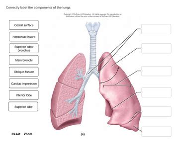
Table of Contents
Correctly Labeling the Components of the Lungs: A Comprehensive Guide
The lungs, the vital organs of respiration, are complex structures with numerous components working in concert to facilitate gas exchange. Understanding the anatomy of the lungs is crucial for healthcare professionals, students, and anyone interested in human physiology. This comprehensive guide will delve into the intricate details of lung anatomy, providing a detailed explanation of each component and offering tips for correctly labeling them. We'll cover everything from the macroscopic structures visible to the naked eye to the microscopic components involved in the critical process of breathing.
The Major Structures of the Lungs: A Macroscopic View
Before diving into the finer details, let's establish a foundational understanding of the major structures of the lungs. These structures are readily identifiable during dissection or with the aid of medical imaging.
1. The Right and Left Lungs: Lobes and Fissures
The lungs are paired organs situated within the thoracic cavity, protected by the rib cage. They are not symmetrical; the right lung is larger and possesses three lobes (superior, middle, and inferior), whereas the left lung is smaller and has only two lobes (superior and inferior) to accommodate the heart. These lobes are separated by fissures:
- Oblique fissure: Present in both lungs, separating the superior and inferior lobes. In the right lung, it also partially separates the middle lobe.
- Horizontal fissure: Found only in the right lung, separating the superior and middle lobes.
Correctly labeling these lobes and fissures is fundamental to understanding lung anatomy. Remember to identify the location of the heart, which influences the shape and size of the left lung.
2. The Trachea and Bronchi: The Airways
Air enters the respiratory system through the trachea (windpipe), a cartilaginous tube that branches into two main bronchi: the right main bronchus and the left main bronchus. These bronchi further subdivide into progressively smaller bronchi, ultimately leading to the microscopic structures responsible for gas exchange. When labeling, pay close attention to the angle of branching: the right main bronchus is shorter, wider, and more vertical than the left, a key anatomical feature.
3. Bronchioles and Alveoli: The Functional Units
The smaller bronchi lead to bronchioles, the smallest conducting airways. These bronchioles terminate in alveolar ducts, which lead to alveolar sacs containing numerous alveoli. Alveoli are the tiny air sacs where gas exchange occurs. Oxygen from inhaled air diffuses into the bloodstream, while carbon dioxide from the blood diffuses into the alveoli to be exhaled. The incredibly large surface area provided by millions of alveoli is essential for efficient gas exchange. Precise labeling requires understanding the hierarchical organization: bronchi → bronchioles → alveolar ducts → alveolar sacs → alveoli.
4. Pulmonary Vessels: Blood Supply
The lungs are richly supplied with blood vessels:
- Pulmonary arteries: Carry deoxygenated blood from the heart to the lungs for oxygenation.
- Pulmonary veins: Carry oxygenated blood from the lungs back to the heart.
- Bronchial arteries and veins: Supply blood to the lung tissue itself, providing nutrients and removing waste products. These vessels are often overlooked but are crucial for the health and functioning of the lung tissue.
Accurately labeling these vessels and understanding their roles in the pulmonary circulation is critical. Remember to differentiate them from the systemic circulation vessels.
Microscopic Anatomy: A Deeper Dive
While the macroscopic structures provide a general overview, a thorough understanding of lung anatomy necessitates examining the microscopic components involved in gas exchange.
1. Alveolar Structure: Type I and Type II Pneumocytes
Alveoli are lined by two types of epithelial cells:
- Type I pneumocytes: These thin, flattened cells form the majority of the alveolar surface area, facilitating efficient gas exchange.
- Type II pneumocytes: These cuboidal cells produce surfactant, a lipoprotein that reduces surface tension within the alveoli, preventing them from collapsing during exhalation.
When labeling microscopic diagrams, clearly distinguish between Type I and Type II pneumocytes and their respective functions.
2. Pulmonary Capillaries: The Gas Exchange Interface
The alveoli are intimately associated with a dense network of pulmonary capillaries. The thin alveolar-capillary membrane facilitates the rapid diffusion of oxygen and carbon dioxide. The close proximity of alveoli and capillaries is a key feature to emphasize when labeling microscopic structures. The thinness of the membrane is essential for efficient gas exchange.
3. Interstitial Tissue: Support and Communication
The alveoli and capillaries are embedded within a delicate interstitial tissue composed of connective tissue, fibroblasts, and immune cells. This tissue provides structural support and facilitates communication between the alveoli and the surrounding structures. While often less visually striking than the alveoli and capillaries, the interstitial tissue plays a vital role in maintaining lung health and function. Accurate labeling should include this often-overlooked component of the lung's microscopic architecture.
Practical Tips for Correct Labeling
Accurately labeling lung components requires careful observation and attention to detail. Here are some practical tips:
- Utilize high-quality anatomical diagrams and models: Refer to reliable sources such as medical textbooks and online resources.
- Start with the macroscopic structures first: Establish a foundational understanding of the lobes, fissures, bronchi, and major vessels before moving to the microscopic level.
- Use clear and concise labels: Avoid ambiguous or overlapping labels.
- Employ color-coding: Different colors can help differentiate various structures and improve visual clarity.
- Practice regularly: Consistent practice is crucial for mastering the labeling of lung components.
- Consult with experts: If you are unsure about specific structures, seek guidance from experienced professionals.
- Use online interactive resources: Many websites offer interactive 3D models of the lungs which allow for exploration and reinforcement of knowledge. These resources can significantly enhance your understanding of spatial relationships between structures.
Clinical Significance of Understanding Lung Anatomy
A thorough understanding of lung anatomy is crucial in various clinical settings:
- Diagnosis and Treatment of Lung Diseases: Accurate identification of affected areas is essential for the diagnosis and treatment of lung diseases such as pneumonia, tuberculosis, lung cancer, and cystic fibrosis. Medical imaging techniques such as X-rays, CT scans, and MRI scans rely heavily on a comprehensive knowledge of lung anatomy for interpretation.
- Surgical Procedures: Thoracic surgeons need precise knowledge of lung anatomy to perform complex procedures such as lung resection and transplantation.
- Respiratory Therapy: Respiratory therapists rely on a detailed understanding of lung anatomy to administer respiratory treatments effectively.
- Medical Research: Researchers involved in pulmonary research need a comprehensive knowledge of lung anatomy to conduct experiments and analyze data correctly.
Conclusion
Correctly labeling the components of the lungs requires a meticulous approach that combines a thorough understanding of macroscopic and microscopic anatomy. By following the guidelines and tips outlined in this guide, you can enhance your knowledge and skills in accurately identifying and labeling the intricate structures of this vital organ. Remember to leverage diverse learning resources, consistent practice, and expert guidance to master this essential aspect of human anatomy. The detailed understanding of lung components is not merely an academic exercise; it's a foundation for crucial advancements in diagnosis, treatment, and research related to respiratory health.
Latest Posts
Latest Posts
-
Compared To Consumer Markets B2b Markets
Mar 20, 2025
-
Identify The Relationship Between The Following Structures
Mar 20, 2025
-
A Successful Quality Strategy Begins With
Mar 20, 2025
-
The Insurance Mechanism Is Based On An Assumption That People
Mar 20, 2025
-
What Is A Proof Of Concept Trying To Achieve
Mar 20, 2025
Related Post
Thank you for visiting our website which covers about Correctly Label The Components Of The Lungs . We hope the information provided has been useful to you. Feel free to contact us if you have any questions or need further assistance. See you next time and don't miss to bookmark.
