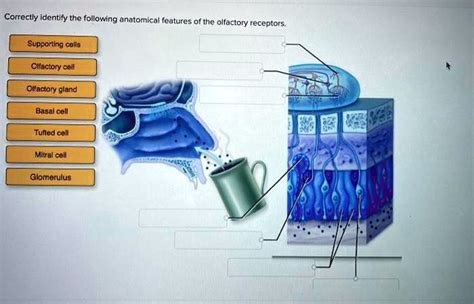
Correctly Identify The Following Anatomical Features Of The Olfactory Receptors.
Holbox
Mar 31, 2025 · 7 min read

Table of Contents
- Correctly Identify The Following Anatomical Features Of The Olfactory Receptors.
- Table of Contents
- Correctly Identify the Following Anatomical Features of the Olfactory Receptors
- The Olfactory Epithelium: The Home of Olfactory Receptors
- 1. Olfactory Receptor Neurons (ORNs): The Sensory Detectives
- 2. Supporting Cells: The Unsung Heroes
- 3. Basal Cells: The Regenerative Force
- Odorant Receptors: The Key to Smell
- Olfactory Transduction: From Odorant to Signal
- The Olfactory Bulb: The First Relay Station
- Higher-Order Olfactory Processing: Perception of Smell
- Clinical Significance and Future Directions
- Latest Posts
- Latest Posts
- Related Post
Correctly Identify the Following Anatomical Features of the Olfactory Receptors
The sense of smell, or olfaction, is a fascinating and complex process, crucial for survival and enriching our daily experiences. At the heart of this process lie the olfactory receptors, specialized sensory neurons responsible for detecting odorant molecules and initiating the signaling cascade that leads to our perception of smell. Understanding the intricate anatomy of these receptors is fundamental to grasping the mechanics of olfaction. This article delves deep into the anatomical features of olfactory receptors, exploring their structure, location, and function in detail.
The Olfactory Epithelium: The Home of Olfactory Receptors
The journey into the anatomy of olfactory receptors begins with their location: the olfactory epithelium. This specialized patch of tissue, approximately 5 cm² in size, resides high within the nasal cavity, on the superior nasal concha and cribriform plate. It's a fascinating microcosm of cellular activity, containing three main cell types crucial for olfaction:
1. Olfactory Receptor Neurons (ORNs): The Sensory Detectives
These are the primary actors in our olfactory experience. ORNs are bipolar neurons, meaning they possess two processes extending from the cell body:
-
Dendrite: This is the receptive portion of the ORN. It extends into the nasal cavity, terminating in a knob-like structure called the olfactory vesicle. From this vesicle, several olfactory cilia project into the mucus layer lining the nasal cavity. These cilia are crucial; they are covered in odorant receptors, the protein molecules that bind to odorant molecules, initiating the transduction process. The high density and large surface area of these cilia maximize the chances of odorant molecule detection. The structure of the cilia itself is remarkably organized, with microtubules and other intracellular components precisely arranged to support the receptors and facilitate signal transduction. Understanding the precise arrangement and composition is crucial for comprehending the sensitivity and specificity of olfactory detection. Mutations or damage to the cilia can lead to significant olfactory impairment.
-
Axon: The other process of the ORN, the axon, travels through the cribriform plate, a bony structure separating the nasal cavity from the brain. These axons collectively form the olfactory nerve (CN I), the first cranial nerve. This nerve carries the olfactory signals to the olfactory bulb, the next stage in the olfactory pathway. The myelination of these axons, while minimal, plays a role in the speed of signal transmission.
2. Supporting Cells: The Unsung Heroes
These are glial cells that provide structural support, metabolic sustenance, and a protective environment for the ORNs. They contribute to the maintenance of the olfactory epithelium's homeostasis, producing mucus and removing debris. Their tight junctions help create the necessary barrier, regulating the access of substances to the olfactory epithelium and protecting against pathogens. The metabolic support they provide is critical for the energy-intensive processes of olfactory transduction and signal transmission. Disruption of the supporting cells' function can compromise the health and function of the ORNs.
3. Basal Cells: The Regenerative Force
These stem cells are located at the base of the olfactory epithelium. They are responsible for the continuous regeneration of ORNs, a unique feature of the olfactory system. ORNs have a relatively short lifespan (around 4-8 weeks), and basal cells provide a constant supply of new neurons to replace the dying ones. This regenerative capacity is essential for maintaining olfactory sensitivity throughout life. Research into basal cells and their mechanisms of differentiation holds promise for developing treatments for olfactory disorders and injuries. Understanding their role in neurogenesis is a critical area of ongoing olfactory research.
Odorant Receptors: The Key to Smell
The remarkable ability to discriminate among thousands of different odorants lies in the diverse array of odorant receptors (ORs) expressed on the olfactory cilia. Each ORN expresses only one type of OR, but the olfactory epithelium as a whole expresses hundreds of different OR genes. This means that different ORNs respond to different odorants, providing a combinatorial code for odor perception.
The ORs are G protein-coupled receptors (GPCRs), a large family of transmembrane receptors involved in a variety of sensory and signaling processes. Their unique structure is characterized by seven transmembrane domains, which allows them to interact with odorant molecules on the extracellular side and initiate intracellular signaling cascades on the intracellular side. The binding specificity of an OR is determined by the amino acid sequence of its transmembrane domains. Slight changes in these sequences can dramatically alter the OR's affinity for particular odorants, contributing to the vast diversity of olfactory sensitivity. The ligand-receptor interaction initiates a series of intracellular events that ultimately lead to the generation of an electrical signal in the ORN.
Olfactory Transduction: From Odorant to Signal
When an odorant molecule binds to its specific OR, it triggers a cascade of events that ultimately converts the chemical signal into an electrical signal. This process, known as olfactory transduction, involves several key steps:
-
Odorant Binding: An odorant molecule binds to its corresponding OR on the olfactory cilia.
-
G-protein Activation: This binding activates a G protein called Golf, initiating a signaling cascade.
-
Adenylate Cyclase Activation: Golf activates adenylate cyclase, an enzyme that converts ATP to cyclic AMP (cAMP).
-
cAMP-gated Ion Channels: cAMP opens cAMP-gated cation channels in the cilia membrane.
-
Depolarization: Influx of sodium and calcium ions depolarizes the ORN membrane, generating a receptor potential.
-
Action Potential Generation: If the depolarization reaches threshold, it triggers action potentials in the ORN axon, transmitting the signal to the olfactory bulb.
The Olfactory Bulb: The First Relay Station
The olfactory nerve fibers carrying signals from the ORNs synapse in the olfactory bulb, a structure located at the base of the frontal lobe of the brain. The olfactory bulb is organized into glomeruli, spherical structures where the axons of ORNs expressing the same type of OR converge. This convergence allows for spatial coding of olfactory information. The glomeruli act as processing centers, integrating signals from multiple ORNs responding to the same odorant. This integrated signal is then transmitted to higher brain regions for further processing and perception. The mitral cells and tufted cells within the glomeruli are the primary output neurons of the olfactory bulb, relaying the olfactory information to various cortical and subcortical areas of the brain. The precise organization and connectivity of the olfactory bulb are critical for the accurate and efficient processing of olfactory information.
Higher-Order Olfactory Processing: Perception of Smell
Signals from the olfactory bulb are transmitted to various brain regions, including the piriform cortex, amygdala, and entorhinal cortex. These areas are involved in different aspects of olfactory processing, such as odor identification, emotional responses to odors, and integration of olfactory information with other sensory modalities.
-
Piriform Cortex: This primary olfactory cortex is involved in odor identification and discrimination. It plays a crucial role in our conscious perception of smell.
-
Amygdala: This area processes the emotional aspects of smell, linking odors to memories and emotions. The amygdala’s role explains why certain smells can evoke strong emotional responses.
-
Entorhinal Cortex: This region is involved in the integration of olfactory information with other sensory modalities, like memory and spatial navigation. The link to memory is crucial to our olfactory experiences.
Clinical Significance and Future Directions
Understanding the anatomy and function of olfactory receptors is essential for diagnosing and treating olfactory disorders. Conditions such as anosmia (loss of smell), hyposmia (reduced smell), and parosmia (distorted smell) can significantly impact a person's quality of life. Damage to the olfactory epithelium, olfactory nerve, or brain areas involved in olfactory processing can cause these disorders. Research into olfactory disorders is crucial for developing effective treatments and interventions. Ongoing research focuses on:
-
Regenerative therapies: Harnessing the regenerative capacity of basal cells to repair damaged olfactory epithelium.
-
Gene therapy: Replacing defective OR genes to restore olfactory function.
-
Drug development: Developing drugs that can protect or restore olfactory function.
-
Neuroimaging techniques: Improving our understanding of olfactory processing in the brain using advanced neuroimaging methods.
In conclusion, the anatomy of olfactory receptors is a marvel of biological engineering, involving a complex interplay of specialized cells, receptors, and brain regions. Understanding this intricate system is critical for appreciating the power and complexity of the sense of smell and its impact on our lives. Continued research will undoubtedly unlock further insights into the mysteries of olfaction, leading to new therapeutic approaches for olfactory disorders and a deeper appreciation for the remarkable sensitivity and specificity of our olfactory system.
Latest Posts
Latest Posts
-
What Is The Free Energy Of Fructose Transport
Apr 04, 2025
-
You Are Filing Documents Into Folders Based On These Criteria
Apr 04, 2025
-
Match Each Of The Following Renal Structures With Their Functions
Apr 04, 2025
-
Match The Cost Variance Component To Its Definition
Apr 04, 2025
-
Correctly Label The Following Functional Regions Of The Cerebral Cortex
Apr 04, 2025
Related Post
Thank you for visiting our website which covers about Correctly Identify The Following Anatomical Features Of The Olfactory Receptors. . We hope the information provided has been useful to you. Feel free to contact us if you have any questions or need further assistance. See you next time and don't miss to bookmark.
