Art-labeling Activity Summary Of Epithelial Tissues
Holbox
Mar 19, 2025 · 6 min read
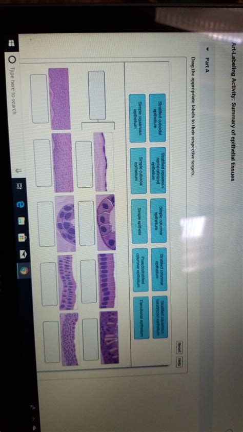
Table of Contents
Art-Labeling Activity Summary of Epithelial Tissues
Epithelial tissues are a fundamental component of the animal body, forming linings, coverings, and glandular structures. Understanding their diverse structures and functions is crucial in various fields, including biology, medicine, and histology. This article provides a comprehensive summary of an art-labeling activity focused on epithelial tissues, detailing their classifications, characteristics, and locations within the body. We will explore different types of epithelial tissue, highlighting their unique features and correlating structure with function. This activity aims to reinforce learning through visual representation and enhance understanding of this complex tissue type.
I. Introduction: The World of Epithelial Tissues
Epithelial tissues, often simply called epithelia, are sheets of tightly packed cells that cover body surfaces, line body cavities and form glands. They are characterized by their cellularity, specialized contacts, polarity, support by connective tissues, avascularity, and regeneration.
A. Key Characteristics of Epithelia:
- Cellularity: Epithelial tissues are composed almost entirely of cells with minimal extracellular matrix.
- Specialized Contacts: Cells are connected by tight junctions, adherens junctions, desmosomes, and gap junctions, ensuring tissue integrity and coordinated function.
- Polarity: Epithelial cells exhibit apical (free) and basal (attached) surfaces, with distinct structural and functional differences.
- Support by Connective Tissues: Epithelial tissues rest on a basement membrane, a layer of extracellular matrix secreted by both epithelial and connective tissue cells. This basement membrane provides structural support and anchors the epithelium.
- Avascularity: Epithelia lack blood vessels; nutrients and waste products are exchanged by diffusion from underlying connective tissues.
- Regeneration: Epithelial tissues have a high regenerative capacity, allowing for rapid repair of damaged areas.
II. Classification of Epithelial Tissues: A Visual Guide
Epithelial tissues are classified based on two primary characteristics: the number of cell layers and the shape of the cells.
A. Number of Cell Layers:
- Simple Epithelium: Composed of a single layer of cells. All cells are in direct contact with the basement membrane. Simple epithelia are typically found where absorption, secretion, or filtration occur.
- Stratified Epithelium: Composed of two or more layers of cells. Only the basal layer is in contact with the basement membrane. Stratified epithelia are typically found in areas subject to wear and tear.
- Pseudostratified Epithelium: Appears stratified but is actually a single layer of cells of varying heights. All cells are in contact with the basement membrane, but their nuclei are at different levels, giving a stratified appearance.
B. Cell Shape:
- Squamous: Flat, scale-like cells.
- Cuboidal: Cube-shaped cells, as wide as they are tall.
- Columnar: Tall, column-shaped cells, taller than they are wide.
- Transitional: Cells that can change shape, typically found in organs that stretch and distend, such as the urinary bladder.
III. Art-Labeling Activity: Detailed Breakdown
The art-labeling activity involves identifying and labeling the different types of epithelial tissues based on their microscopic images. This exercise strengthens understanding by associating visual representations with their structural and functional properties. The activity may include images depicting:
A. Simple Squamous Epithelium:
- Location: Lining of blood vessels (endothelium), body cavities (mesothelium), alveoli of lungs.
- Function: Diffusion, filtration, secretion.
- Image Characteristics: Thin, flat cells; nuclei appear as flattened ovals.
Labeling Points: Identify the single layer of flat cells, the basement membrane, and the location where this epithelium is found. Note the thinness of the cells, facilitating rapid diffusion.
B. Simple Cuboidal Epithelium:
- Location: Kidney tubules, ducts of glands, surface of ovaries.
- Function: Secretion, absorption.
- Image Characteristics: Cube-shaped cells; round, centrally located nuclei.
Labeling Points: Identify the single layer of cube-shaped cells, the basement membrane, and the centrally located nuclei. Note the presence of secretory granules or microvilli depending on the specific location.
C. Simple Columnar Epithelium:
- Location: Lining of digestive tract (stomach to rectum), gallbladder.
- Function: Secretion, absorption, protection.
- Image Characteristics: Tall, column-shaped cells; oval nuclei usually located near the base of the cells. May contain goblet cells (mucus-secreting) or microvilli (for absorption).
Labeling Points: Identify the single layer of tall cells, the basement membrane, the location of the nuclei, and the presence of goblet cells or microvilli if present. Highlight the relationship between cell structure and absorption or secretion.
D. Pseudostratified Columnar Epithelium:
- Location: Lining of trachea, bronchi, and parts of the male reproductive system.
- Function: Secretion (mucus), propulsion of mucus by cilia.
- Image Characteristics: Single layer of cells of varying heights; all cells contact the basement membrane; nuclei at different levels; often ciliated.
Labeling Points: Identify the single layer of cells, the basement membrane, the varying heights of the cells, and the presence of cilia. Explain why it appears stratified despite being a single layer. Highlight the function of cilia in mucus movement.
E. Stratified Squamous Epithelium:
- Location: Epidermis of skin, lining of esophagus, mouth, and vagina.
- Function: Protection against abrasion, dehydration, and infection.
- Image Characteristics: Many cell layers; apical cells are flat, while basal cells may be cuboidal or columnar.
Labeling Points: Identify the multiple layers of cells, the flat apical cells, and the deeper cuboidal or columnar basal cells. Explain how the structure contributes to its protective function. Distinguish between keratinized (skin) and non-keratinized (esophagus) varieties.
F. Stratified Cuboidal Epithelium:
- Location: Ducts of sweat glands and salivary glands.
- Function: Protection, secretion.
- Image Characteristics: Two or more layers of cube-shaped cells.
Labeling Points: Identify the multiple layers of cube-shaped cells and the location of their nuclei. Explain the function of this epithelium in the context of its location.
G. Stratified Columnar Epithelium:
- Location: Male urethra, parts of the pharynx.
- Function: Protection, secretion.
- Image Characteristics: Several layers of cells; superficial cells are columnar, while basal cells may vary in shape.
Labeling Points: Identify the multiple layers of cells, the columnar apical cells, and the varying shapes of basal cells. Explain its limited distribution and functions.
H. Transitional Epithelium:
- Location: Lining of urinary bladder, ureters, and parts of the urethra.
- Function: Stretching and distension.
- Image Characteristics: Cells change shape depending on the degree of distension; relaxed state shows dome-shaped cells, while stretched state shows flattened cells.
Labeling Points: Identify the ability of cells to change shape and explain how this allows for distension and contraction of the urinary bladder.
IV. Correlation of Structure and Function: A Deeper Dive
The art-labeling activity should emphasize the correlation between the structure of epithelial tissues and their functions. For instance:
- Simple squamous epithelium's thinness allows for efficient diffusion and filtration in the lungs and blood vessels.
- Simple cuboidal epithelium's cube shape provides ample cytoplasm for secretion and absorption in glands and kidney tubules.
- Simple columnar epithelium's height allows for increased surface area for absorption and secretion in the digestive tract. The presence of microvilli further enhances absorption.
- Stratified squamous epithelium's multiple layers provide protection against abrasion and dehydration in the skin and other areas prone to friction.
- Transitional epithelium's ability to change shape allows it to accommodate the changing volume of urine in the urinary bladder.
V. Conclusion: Beyond the Label
The art-labeling activity serves as a valuable tool for understanding the intricacies of epithelial tissues. By visually identifying and labeling these tissues, students strengthen their understanding of their diverse structures and functions. However, the activity should not end with simple labeling. It should stimulate critical thinking and lead to a deeper appreciation of how the structure of these tissues directly relates to their roles in maintaining homeostasis within the body. This understanding is essential for comprehending a broad range of physiological processes and pathophysiological conditions. Further exploration into the specific functions of different epithelial tissues in different organ systems would greatly enhance learning and retention.
Latest Posts
Latest Posts
-
Real Time Physics Lab 7 Homework Answers
Mar 19, 2025
-
What Do Sutures Gomphoses And Syndesmoses Have In Common
Mar 19, 2025
-
In Order To Promote Growth In Living Standards Policymakers Must
Mar 19, 2025
-
Suppose That A Small Town Wants To Install Street Lamps
Mar 19, 2025
-
What Is Something An Excellent Business Writer Would Do
Mar 19, 2025
Related Post
Thank you for visiting our website which covers about Art-labeling Activity Summary Of Epithelial Tissues . We hope the information provided has been useful to you. Feel free to contact us if you have any questions or need further assistance. See you next time and don't miss to bookmark.
