An Indicator Of An Expanding Intracranial Hematoma
Holbox
Mar 19, 2025 · 6 min read
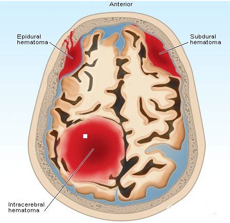
Table of Contents
An Indicator of an Expanding Intracranial Hematoma: Recognizing the Critical Signs
An expanding intracranial hematoma represents a life-threatening neurosurgical emergency. Early detection and intervention are crucial for improving patient outcomes and survival. This article will delve into the multifaceted indicators of an expanding intracranial hematoma, emphasizing the critical signs and symptoms that necessitate immediate medical attention. Understanding these indicators empowers healthcare professionals and caregivers to act swiftly, potentially saving lives.
Understanding Intracranial Hematomas
Before examining the indicators of expansion, let's briefly review intracranial hematomas themselves. These are collections of blood within the skull, typically resulting from trauma (e.g., head injuries, falls, motor vehicle accidents) or spontaneous bleeding (e.g., aneurysms, arteriovenous malformations). Several types exist, each with its own characteristics and locations:
-
Epidural Hematoma: Located between the skull and the dura mater (the outermost layer of the brain's protective coverings). These often result from arterial bleeding and expand rapidly.
-
Subdural Hematoma: Situated between the dura mater and the arachnoid mater (the middle layer). Usually venous in origin, they can expand more slowly than epidural hematomas, but still pose a significant threat.
-
Subarachnoid Hemorrhage: Bleeding into the subarachnoid space, the area between the arachnoid mater and the pia mater (the innermost layer). This is often associated with aneurysmal rupture.
-
Intracerebral Hematoma: Bleeding directly within the brain tissue itself. This can arise from trauma, hypertension, or other vascular disorders.
The critical aspect is that any of these hematomas can expand, increasing intracranial pressure (ICP) and causing devastating neurological consequences.
Key Indicators of an Expanding Intracranial Hematoma
The signs and symptoms of an expanding intracranial hematoma are often subtle initially, making early detection challenging. However, a deteriorating neurological status should always raise suspicion. The key indicators can be broadly categorized as:
1. Changes in Level of Consciousness (LOC):
This is arguably the most crucial indicator. Deterioration in LOC, progressing from alertness to drowsiness, confusion, lethargy, or even coma, strongly suggests an expanding hematoma. This is because the increasing ICP compresses brain tissue, impairing its function.
- Early signs: Slight confusion, difficulty concentrating, slowed responses.
- Late signs: Unresponsiveness, loss of consciousness, inability to be aroused.
Careful and continuous monitoring of LOC is paramount. Any change, no matter how small, warrants immediate medical assessment.
2. Neurological Deficit Progression:
As the hematoma expands, it exerts pressure on specific brain regions, leading to a worsening of neurological deficits. These deficits can manifest in various ways, depending on the location of the hematoma:
- Hemiparesis/Hemiplegia: Weakness or paralysis on one side of the body.
- Facial Palsy: Drooping on one side of the face.
- Aphasia: Difficulty with speech or understanding language.
- Ataxia: Loss of coordination and balance.
- Visual Field Defects: Loss of vision in part of the visual field.
- Pupillary Changes: Unequal pupil size (anisocoria) or sluggish pupillary response to light. Unilateral dilated and fixed pupil is a particularly ominous sign indicating significant brainstem compression.
The progression of these deficits, even if initially mild, is a major warning sign. For instance, a patient who initially experiences slight weakness on one side of the body and then develops complete paralysis needs urgent intervention.
3. Headache:
While headaches are common, a sudden onset of severe headache, particularly if accompanied by other neurological symptoms, is highly suggestive of an intracranial hematoma. The headache may be described as the "worst headache of my life." The intensity and character of the headache might change over time as the hematoma expands.
4. Vomiting:
Projectile vomiting without nausea can be a sign of increased intracranial pressure. This occurs because the pressure stimulates the vomiting center in the brainstem.
5. Altered Respiratory Pattern:
Changes in breathing pattern, such as Cheyne-Stokes respiration (alternating periods of apnea and hyperpnea) or irregular breathing, can signify brainstem compression due to rising ICP. This indicates a critically deteriorating situation.
6. Hypertension and Bradycardia (Cushing's Triad):
Cushing's triad is a late and ominous sign of increased intracranial pressure. It consists of:
- Hypertension: Elevated blood pressure with a widening pulse pressure (the difference between systolic and diastolic pressure).
- Bradycardia: Slow heart rate.
- Irregular Respirations: Abnormal breathing patterns.
The appearance of Cushing's triad indicates severe brainstem compression and signifies impending herniation, a life-threatening condition where brain tissue is squeezed through the openings in the skull.
7. Decreasing Level of Responsiveness:
As ICP rises, the brain's ability to function properly decreases, leading to a gradual decline in responsiveness. This can manifest as slowed responses to commands, difficulty following instructions, or decreased alertness. This is a particularly concerning sign, indicating a potential rapid expansion of the hematoma.
8. Post-traumatic Changes:
In cases of traumatic brain injury, worsening of initial symptoms such as headache, confusion, or dizziness after an initial period of improvement is a significant warning sign. This "second impact" suggests hematoma expansion.
Imaging Studies: Confirming the Diagnosis
While the clinical signs mentioned above are crucial for suspicion, imaging studies are essential for confirming the diagnosis and assessing the size and location of the hematoma. The most common imaging techniques used are:
-
Computed Tomography (CT) Scan: A rapid and readily available imaging method that provides excellent visualization of intracranial structures. CT scans are particularly useful in the acute setting to identify intracranial bleeding.
-
Magnetic Resonance Imaging (MRI): Provides more detailed images than CT scans, particularly for visualizing soft tissues. MRI is often used for follow-up imaging after the initial diagnosis.
Management of Expanding Intracranial Hematomas
The management of expanding intracranial hematomas is primarily surgical. The goal is to evacuate the hematoma, relieve the pressure on the brain, and improve neurological function. The specific surgical approach depends on the type and location of the hematoma. In addition to surgery, supportive measures such as:
- Intravenous Fluids: Maintain adequate hydration.
- Blood Pressure Control: Manage hypertension.
- Respiratory Support: Assist with breathing if needed.
- Monitoring: Close monitoring of vital signs, neurological status, and ICP.
Conclusion: Prognosis and Prevention
The prognosis for patients with expanding intracranial hematomas depends on several factors, including the size and location of the hematoma, the rapidity of expansion, and the patient's overall health. Early detection and prompt surgical intervention are crucial for improving outcomes. While many cases result from trauma or spontaneous bleeding, some risk factors can be modified:
- Control of Hypertension: High blood pressure significantly increases the risk of intracerebral hemorrhage.
- Anticoagulation Management: Careful monitoring and management of anticoagulant medications are essential to minimize the risk of bleeding.
- Alcohol Avoidance: Excessive alcohol consumption is associated with an increased risk of intracranial bleeding.
Prompt recognition of the indicators of an expanding intracranial hematoma is paramount. Immediate medical attention is crucial for initiating appropriate treatment and improving the chances of a favorable outcome. The combination of clinical awareness, rapid diagnostic imaging, and prompt neurosurgical intervention can make the difference between life and death. This underscores the importance of recognizing and acting upon the warning signs described above. Remember, timely intervention is key to improving patient survival and neurological recovery.
Latest Posts
Latest Posts
-
How Many Times Does 15 Go Into 135
Mar 20, 2025
-
The Viral Infection Hepatitis A Can Be Most Effectively
Mar 20, 2025
-
Stagflation Occurs When High Inflation Combines With
Mar 20, 2025
-
A Radio Station Is Giving Away Tickets To A Play
Mar 20, 2025
-
Reading The World Ideas That Matter 4th Edition Michael Austin
Mar 20, 2025
Related Post
Thank you for visiting our website which covers about An Indicator Of An Expanding Intracranial Hematoma . We hope the information provided has been useful to you. Feel free to contact us if you have any questions or need further assistance. See you next time and don't miss to bookmark.
