Where In The Neuron Is An Action Potential Initially Generated
Holbox
Mar 13, 2025 · 6 min read
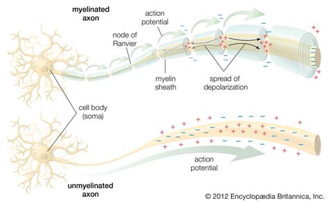
Table of Contents
- Where In The Neuron Is An Action Potential Initially Generated
- Table of Contents
- Where in the Neuron is an Action Potential Initially Generated?
- The Neuron: A Brief Overview
- The Role of the Axon Hillock in Action Potential Initiation
- 1. High Density of Voltage-Gated Sodium Channels:
- 2. Spatial Summation and Temporal Summation:
- 3. Low Membrane Resistance:
- 4. Absence of Myelin Sheath:
- The Process of Action Potential Initiation
- Why the Axon Hillock and Not Other Regions?
- Variations and Exceptions
- Conclusion
- Latest Posts
- Latest Posts
- Related Post
Where in the Neuron is an Action Potential Initially Generated?
The human nervous system, a marvel of biological engineering, relies on the rapid transmission of electrical signals known as action potentials. These fleeting bursts of electrical activity are the fundamental units of neural communication, allowing us to perceive the world, control our movements, and think. But where does this crucial process begin? Understanding the precise location of action potential initiation is key to understanding how our nervous system functions. This article delves deep into the intricacies of neuronal structure and the biophysics of action potential generation, ultimately answering the question: where, precisely, in the neuron does an action potential initially arise?
The Neuron: A Brief Overview
Before diving into the specifics of action potential generation, a brief overview of neuronal structure is essential. Neurons, the fundamental units of the nervous system, are highly specialized cells designed for rapid communication. They possess several key components:
- Dendrites: These branching extensions receive signals from other neurons. They are studded with receptors that bind neurotransmitters, the chemical messengers that transmit signals across the synapse, the junction between two neurons.
- Soma (Cell Body): The soma contains the nucleus and other essential organelles, responsible for the neuron's metabolic processes. It integrates the signals received from dendrites.
- Axon Hillock: This specialized region of the neuron, located at the junction between the soma and the axon, plays a crucial role in action potential initiation. It acts as a trigger zone.
- Axon: The axon is a long, slender projection that transmits the action potential away from the soma to other neurons, muscles, or glands.
- Axon Terminals (Synaptic Terminals): At the end of the axon, these structures release neurotransmitters to communicate with other cells.
The Role of the Axon Hillock in Action Potential Initiation
While the dendrites and soma receive and process incoming signals, the axon hillock is the critical region where the decision to generate an action potential is made. This isn't a random location; it's strategically positioned due to its unique biophysical properties. Several factors contribute to its role as the action potential initiation site:
1. High Density of Voltage-Gated Sodium Channels:
The axon hillock has a significantly higher density of voltage-gated sodium (Na+) channels compared to other parts of the neuron, especially the soma and dendrites. These channels are crucial for action potential generation. They are normally closed, but when the membrane potential depolarizes (becomes less negative) to a certain threshold, they open rapidly, allowing a large influx of Na+ ions into the neuron. This rapid influx of positive charge is what drives the rapid depolarization phase of the action potential. The high concentration of these channels in the axon hillock ensures that a sufficient depolarization can trigger the opening of enough channels to reach the threshold for action potential generation.
2. Spatial Summation and Temporal Summation:
The axon hillock acts as an integrative center, receiving signals from multiple dendrites. This allows for both spatial summation (summing of signals from different dendrites) and temporal summation (summing of signals arriving at the same dendrite in close succession). If the summed excitatory postsynaptic potentials (EPSPs) – which are depolarizations – reach the threshold potential at the axon hillock, an action potential is triggered.
3. Low Membrane Resistance:
The axon hillock also exhibits a relatively low membrane resistance. This means that the current flowing into the axon hillock from the soma and dendrites is less likely to leak out through the membrane before reaching the threshold for voltage-gated sodium channel activation. This efficient current flow ensures that even relatively small depolarizations can effectively trigger an action potential.
4. Absence of Myelin Sheath:
Unlike much of the axon, the axon hillock is typically unmyelinated. The myelin sheath, a fatty insulating layer surrounding many axons, speeds up action potential conduction. However, its absence at the axon hillock is critical for action potential initiation. The unmyelinated nature ensures that the voltage changes are not buffered or reduced as they would be in a myelinated section, allowing for accurate threshold detection and action potential initiation.
The Process of Action Potential Initiation
Let's trace the steps involved in action potential initiation at the axon hillock:
-
Synaptic Input: Neurotransmitters released from presynaptic neurons bind to receptors on the dendrites, triggering either EPSPs (excitatory postsynaptic potentials) or IPSPs (inhibitory postsynaptic potentials). EPSPs depolarize the membrane, while IPSPs hyperpolarize it.
-
Signal Integration at the Soma: The EPSPs and IPSPs propagate passively towards the axon hillock, undergoing both spatial and temporal summation.
-
Threshold Reaching: If the summed depolarization at the axon hillock reaches the threshold potential (typically around -55 mV), it triggers the opening of voltage-gated sodium channels.
-
Rapid Depolarization: The rapid influx of sodium ions causes a dramatic depolarization of the membrane, reaching a peak of around +30 mV. This is the rising phase of the action potential.
-
Repolarization: Voltage-gated potassium (K+) channels then open, allowing potassium ions to flow out of the neuron. This efflux of positive charge repolarizes the membrane, returning it to its resting potential.
-
Hyperpolarization: The potassium channels remain open slightly longer than necessary, leading to a brief period of hyperpolarization (membrane potential becomes more negative than the resting potential).
-
Return to Resting Potential: Finally, ion pumps restore the ion gradients across the membrane, bringing the membrane potential back to its resting state, ready to receive new signals.
Why the Axon Hillock and Not Other Regions?
The strategic positioning and unique properties of the axon hillock make it ideally suited for initiating action potentials. Why not other regions?
- Dendrites: Dendrites receive signals but lack the high density of voltage-gated sodium channels needed to generate the rapid depolarization required for an action potential.
- Soma: While the soma integrates signals, its lower density of voltage-gated sodium channels makes it less efficient at initiating action potentials compared to the axon hillock. The soma's broader diameter also increases the membrane resistance, hindering the efficient spread of depolarization.
- Axon: The axon's primary role is to conduct, not initiate, action potentials. The myelinated sections of the axon further reduce the likelihood of action potential initiation.
The axon hillock, therefore, acts as a crucial decision-making point, ensuring that an action potential is generated only when the summed input from various dendritic branches reaches a sufficient level. This mechanism prevents spurious action potentials and ensures efficient and controlled neuronal communication.
Variations and Exceptions
While the axon hillock is the primary site of action potential initiation in many neurons, there are exceptions and variations depending on the neuronal type and its specific function. Some neurons may have multiple initiation sites, or the initiation site might be slightly displaced from the classic axon hillock location. Nevertheless, the underlying principles of high density voltage-gated sodium channels, low membrane resistance, and effective signal integration remain crucial for action potential initiation, regardless of the precise location.
Conclusion
The initiation of an action potential is a carefully orchestrated process, critically dependent on the unique properties of the axon hillock. Its high concentration of voltage-gated sodium channels, low membrane resistance, and strategic location facilitate the summation of synaptic inputs and the efficient generation of the all-or-nothing electrical signal that underpins neural communication. Understanding the role of the axon hillock in action potential initiation provides crucial insight into the fundamental mechanisms that govern the operation of our nervous system. This knowledge is essential for research in neurobiology, neuroscience, and related fields, potentially leading to better treatments for neurological disorders and a deeper understanding of the human brain.
Latest Posts
Latest Posts
-
How Many Pounds Is 105 Kilograms
May 19, 2025
-
How Many Oz In 650 Ml
May 19, 2025
-
How Far Is A 1000 Feet
May 19, 2025
-
How Many Feet Is 73 Inches
May 19, 2025
-
How Many Feet Is 46 Inches
May 19, 2025
Related Post
Thank you for visiting our website which covers about Where In The Neuron Is An Action Potential Initially Generated . We hope the information provided has been useful to you. Feel free to contact us if you have any questions or need further assistance. See you next time and don't miss to bookmark.