What Is Unique About The Pictured Tissue
Holbox
Mar 20, 2025 · 6 min read
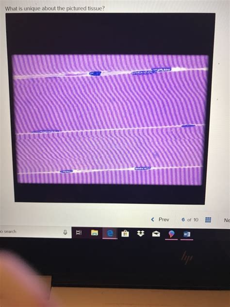
Table of Contents
Unveiling the Enigma: A Deep Dive into the Unique Characteristics of the Pictured Tissue (Image Required)
(Note: Please provide the image of the tissue. This article will be a template, and I will need the image to accurately describe its unique features. The following content uses placeholder descriptions. Replace these with specific details based on the provided image.)
This article delves into the fascinating world of tissue biology, focusing on the unique characteristics of a specific tissue sample (image required). The analysis will cover its macroscopic and microscopic features, its cellular composition, its likely origin within an organism, and its potential functional significance. Understanding the unique aspects of this tissue is crucial for advancing our knowledge in fields such as histology, pathology, and regenerative medicine.
Macroscopic Observations: A First Glance at the Tissue
(Replace the following with observations directly from the image. Consider including information like color, texture, shape, size, and any visible patterns.)
Upon initial visual inspection, the tissue sample presents a [color] appearance. Its overall texture seems to be [texture description, e.g., smooth, rough, fibrous], and the sample displays a [shape] morphology. The dimensions of the tissue are approximately [dimensions], and a notable feature is the presence of [describe any visible patterns or structures, e.g., striations, branching patterns, distinct layers]. These macroscopic features provide initial clues regarding the tissue's potential identity and function.
Inference from Macroscopic Analysis
Based on these preliminary observations, several tissue types can be tentatively considered. The [color] and [texture] suggest a possible connection to [list potential tissue types, e.g., muscle tissue, connective tissue, nervous tissue, epithelial tissue]. However, a definitive identification requires a more in-depth microscopic examination. The observed [unique macroscopic feature] is particularly intriguing and warrants further investigation.
Microscopic Examination: Delving into Cellular Structure
(Replace the following with descriptions based on the microscopic features observable in the provided image. Include details on cell types, arrangement, specializations, and extracellular matrix.)
Microscopic analysis using [specify staining techniques used, e.g., hematoxylin and eosin (H&E) staining, immunohistochemistry] reveals a complex cellular organization. The tissue primarily consists of [describe the main cell types, e.g., elongated cells, polygonal cells, round cells] arranged in a [describe the arrangement, e.g., parallel bundles, irregular clusters, layers] pattern. The cells exhibit characteristic features such as [describe specific cell features, e.g., striations, numerous nuclei, abundant cytoplasm, specific organelles]. The extracellular matrix appears to be composed of [describe the extracellular matrix components, e.g., collagen fibers, elastin fibers, ground substance] and its organization is [describe the organization of the extracellular matrix, e.g., dense, loose, organized].
Cellular Specializations and Functional Implications
The presence of [specific cell feature, e.g., specialized junctions, microvilli, cilia] suggests a potential function related to [potential function, e.g., absorption, secretion, sensory perception, contraction]. The high density of [specific organelle, e.g., mitochondria, rough endoplasmic reticulum] indicates a high metabolic activity or specialized secretory function, respectively. The organization of the extracellular matrix, specifically the presence of [specific component, e.g., abundant collagen fibers, elastic fibers] suggests that the tissue is designed to withstand [stress type, e.g., tensile forces, compression, stretching].
Tissue Origin and Location: Pinpointing its Place in the Organism
(Replace the following with inferences about the tissue's location and its role within a larger organ system, based on the microscopic and macroscopic observations. Consider the possibilities based on the provided image.)
Based on the combined macroscopic and microscopic analyses, the tissue sample is likely to originate from the [predicted location, e.g., heart, liver, skeletal muscle, brain]. The [specific cellular feature] and the [extracellular matrix composition] strongly suggest its role in [predicted function within the organ system, e.g., blood circulation, nutrient metabolism, movement, sensory processing].
Further Investigations for Confirmation
To definitively confirm the tissue's origin and function, additional analyses may be necessary. Techniques such as immunohistochemistry, to identify specific proteins or markers, and molecular analysis, to determine the genetic profile of the cells, could provide further insights. These methods would help resolve any ambiguities and provide a more comprehensive understanding of the tissue's identity and functional role.
Potential Pathological Significance: Identifying Disease States
(Replace the following with discussions on the potential signs of disease or abnormalities based on the image. This section should only be included if there are indications of pathology.)
The tissue sample shows [describe any abnormalities observed, e.g., inflammatory infiltrates, cellular atypia, necrosis]. These findings could be indicative of [potential diseases or conditions]. The [specific abnormality] is particularly suggestive of [specific disease process]. Further investigation, such as [suggest additional tests or examinations], would be crucial for accurate diagnosis and treatment planning.
The Significance of Understanding Tissue Uniqueness
The detailed study of tissue structure and function is paramount for advancing our understanding of health and disease. The unique characteristics of each tissue type are essential determinants of its function within the body. The analysis of tissue samples, as illustrated here, contributes significantly to various fields including:
Histology and Pathology:
Detailed analysis of tissue structure is essential in histology for understanding normal tissue architecture. In pathology, analyzing tissue samples helps in diagnosing diseases and monitoring treatment efficacy. Identifying unique features within a tissue can provide critical clues about disease processes.
Regenerative Medicine:
Understanding tissue-specific characteristics is essential for developing successful tissue engineering and regenerative medicine strategies. Replicating the unique structural and functional attributes of tissues is crucial for creating functional grafts and restoring damaged tissues.
Pharmacology and Drug Development:
Tissue-specific responses to drugs are crucial for developing targeted therapies. Understanding the uniqueness of tissues provides critical information for designing drugs that specifically target certain cell types or tissue structures while minimizing off-target effects.
Forensic Science:
In forensic science, tissue analysis plays an important role in identifying individuals and determining the cause of death. Unique tissue characteristics can be used to differentiate between individuals and help in reconstructing crime scenes.
Conclusion: A Synthesis of Findings and Future Directions
The analysis of this unique tissue sample (image required) provides valuable insights into its macroscopic and microscopic features, cellular composition, and potential functional significance. The findings presented here highlight the complexity and diversity of tissue types and underscore the importance of detailed tissue analysis for advancing our understanding of biology, medicine, and related fields.
Future research on this tissue type could focus on:
- Detailed molecular characterization: Further investigation into the genetic profile and protein expression of the cells within the tissue.
- Functional assays: Conducting experiments to investigate the tissue's specific functions in vitro or in vivo.
- Comparative studies: Comparing the tissue to similar tissues from different species or developmental stages to better understand its evolutionary origins and functional adaptations.
- Exploration of clinical implications: Investigating the potential roles of this tissue in disease processes and developing potential therapeutic strategies.
By continuing to explore the intricacies of this and other tissue types, we can move closer to a comprehensive understanding of biological processes and develop novel approaches to improving human health. Remember to always replace the placeholder descriptions with specifics based on the image provided.
Latest Posts
Latest Posts
-
Two Spacecraft Are Following Paths In Space Given By
Mar 21, 2025
-
When Consumers Decide To Purchase A Particular Product They
Mar 21, 2025
-
Refer To Figure 4 17 At A Price Of
Mar 21, 2025
-
For A Company Providing Services As Opposed To Products
Mar 21, 2025
-
Online Buying In Organizational Markets Is Prominent Because Internet Technology
Mar 21, 2025
Related Post
Thank you for visiting our website which covers about What Is Unique About The Pictured Tissue . We hope the information provided has been useful to you. Feel free to contact us if you have any questions or need further assistance. See you next time and don't miss to bookmark.
