The Juxtaglomerular Apparatus Regulates The Filtration Rate By
Holbox
Mar 27, 2025 · 5 min read
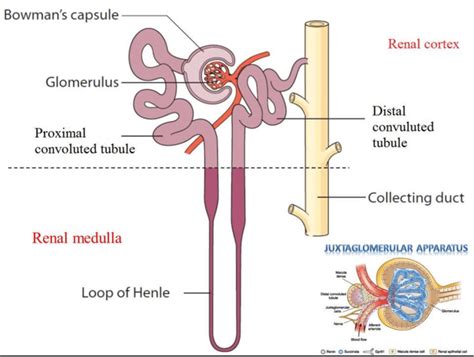
Table of Contents
- The Juxtaglomerular Apparatus Regulates The Filtration Rate By
- Table of Contents
- The Juxtaglomerular Apparatus: Regulating Glomerular Filtration Rate (GFR)
- Understanding the Juxtaglomerular Apparatus
- 1. Granular Cells (Juxtaglomerular Cells):
- 2. Macula Densa:
- 3. Extraglomerular Mesangial Cells:
- Mechanisms of GFR Regulation by the JGA
- 1. The Renin-Angiotensin-Aldosterone System (RAAS):
- 2. Tubuloglomerular Feedback (TGF):
- 3. Myogenic Mechanism:
- 4. Neural Regulation:
- Clinical Significance of JGA Dysfunction
- Further Research and Future Directions
- Conclusion
- Latest Posts
- Latest Posts
- Related Post
The Juxtaglomerular Apparatus: Regulating Glomerular Filtration Rate (GFR)
The precise control of glomerular filtration rate (GFR) is paramount for maintaining homeostasis within the human body. Too high a GFR leads to excessive fluid and solute loss, while too low a GFR results in inadequate waste removal and fluid retention. This delicate balancing act is largely orchestrated by the juxtaglomerular apparatus (JGA), a specialized structure located within the kidney's nephron. This article will delve deep into the intricate mechanisms by which the JGA regulates GFR, exploring its components, their functions, and the interplay of hormonal and neural influences.
Understanding the Juxtaglomerular Apparatus
The JGA is a remarkable micro-anatomical complex formed by the interaction of the distal convoluted tubule (DCT) and the afferent arteriole. It's not a discrete organ, but rather a specialized region where these two structures come into close proximity. The key cellular components of the JGA include:
1. Granular Cells (Juxtaglomerular Cells):
Located within the walls of the afferent arteriole, these modified smooth muscle cells are the primary source of renin, a crucial hormone in the renin-angiotensin-aldosterone system (RAAS). These cells are mechanoreceptors, meaning they are sensitive to changes in blood pressure within the afferent arteriole. A decrease in blood pressure stimulates renin release.
2. Macula Densa:
This specialized group of epithelial cells resides in the distal convoluted tubule (DCT), where it sits adjacent to the afferent arteriole. The macula densa cells act as chemoreceptors, detecting changes in the concentration of sodium chloride (NaCl) in the tubular fluid. A decrease in NaCl concentration signals a decrease in GFR, prompting the macula densa to release signals that ultimately lead to increased renin secretion.
3. Extraglomerular Mesangial Cells:
These cells are located between the afferent and efferent arterioles and connect the granular cells and the macula densa. Their function is not fully understood, but they are believed to play a role in conveying signals between the macula densa and granular cells and potentially influencing the activity of both. They may also contribute to the regulation of glomerular filtration pressure.
Mechanisms of GFR Regulation by the JGA
The JGA's regulation of GFR is a complex interplay of various feedback loops involving both local and systemic factors. The primary mechanism centers around the renin-angiotensin-aldosterone system (RAAS), but other factors also contribute significantly.
1. The Renin-Angiotensin-Aldosterone System (RAAS):
The RAAS is the cornerstone of JGA's influence on GFR. Let's break down the process:
- Renin Release: Triggered by a decrease in blood pressure (detected by granular cells), a decrease in NaCl concentration in the DCT (detected by macula densa), or sympathetic nervous system activation.
- Angiotensinogen Conversion: Renin converts angiotensinogen (a liver-produced protein) into angiotensin I.
- Angiotensin I to Angiotensin II: Angiotensin-converting enzyme (ACE), primarily found in the lungs, converts angiotensin I into angiotensin II, a potent vasoconstrictor.
- Vasoconstriction: Angiotensin II constricts the efferent arteriole, increasing glomerular hydrostatic pressure and thus maintaining GFR despite the initial decrease in blood pressure. This selective constriction is crucial; constriction of the afferent arteriole would drastically reduce GFR.
- Aldosterone Release: Angiotensin II also stimulates the adrenal cortex to release aldosterone.
- Sodium and Water Retention: Aldosterone promotes sodium reabsorption in the distal convoluted tubule and collecting duct, leading to increased water reabsorption, expanding blood volume, and ultimately increasing blood pressure.
This entire process effectively restores GFR to normal levels by increasing blood pressure and blood volume.
2. Tubuloglomerular Feedback (TGF):
The macula densa's role in monitoring NaCl concentration is vital in the tubuloglomerular feedback (TGF) mechanism. When GFR increases, more NaCl reaches the macula densa. This triggers a paracrine signaling cascade that leads to:
- Afferent Arteriole Constriction: The macula densa signals the afferent arteriole to constrict, decreasing blood flow into the glomerulus and reducing GFR.
- Mesangial Cell Contraction: The macula densa may also influence mesangial cell contraction, reducing the glomerular filtration surface area and further decreasing GFR.
This negative feedback loop ensures that GFR remains within a physiological range despite fluctuations in systemic blood pressure.
3. Myogenic Mechanism:
The afferent arteriole's smooth muscle cells possess inherent myogenic properties; they respond directly to changes in pressure. An increase in blood pressure stretches the afferent arteriole, triggering vasoconstriction and reducing GFR. Conversely, a decrease in blood pressure causes vasodilation, maintaining GFR to some extent.
4. Neural Regulation:
The sympathetic nervous system exerts significant control over GFR. During stress or reduced blood volume, sympathetic nerve activation:
- Increases Renin Release: Stimulates granular cells to release more renin, initiating the RAAS cascade.
- Constricts Afferent Arteriole: Directly constricts the afferent arteriole, reducing GFR.
This neural influence is crucial for maintaining blood pressure and perfusion to vital organs during emergencies.
Clinical Significance of JGA Dysfunction
Dysregulation of the JGA can lead to several clinical conditions:
- Hypertension: Excessive renin production can cause increased angiotensin II levels, leading to persistent vasoconstriction and hypertension.
- Hypotension: Insufficient renin production can result in reduced blood volume and hypotension.
- Kidney Failure: Damage to the JGA can impair its ability to regulate GFR, leading to impaired kidney function and ultimately kidney failure.
- Fluid and Electrolyte Imbalances: Impaired JGA function can cause disturbances in sodium, potassium, and water balance.
Further Research and Future Directions
While significant strides have been made in understanding the JGA's role in GFR regulation, several aspects remain to be fully elucidated. Further research is needed to fully understand:
- The precise signaling pathways between the macula densa and granular cells.
- The complete role of extraglomerular mesangial cells in GFR regulation.
- The interaction between the JGA and other renal regulatory mechanisms.
- The development of novel therapeutic strategies targeting the JGA to treat hypertension and kidney diseases.
Conclusion
The juxtaglomerular apparatus is a sophisticated regulatory system vital for maintaining a stable glomerular filtration rate. Its intricate interplay of hormonal, neural, and local feedback mechanisms ensures that the kidneys can efficiently filter blood, remove waste products, and maintain fluid and electrolyte balance. Understanding the complex processes involved in JGA function is essential for diagnosing and treating various renal and cardiovascular disorders. Continued research will undoubtedly unveil further intricacies of this crucial physiological system. The remarkable precision and adaptability of the JGA underscore its importance in overall body homeostasis. Future advances in our knowledge will certainly lead to more effective interventions in the treatment of various kidney and cardiovascular diseases.
Latest Posts
Latest Posts
-
Shortly After Assisting A 65 Year Old
Mar 30, 2025
-
What Are The Potential People Change Issues Facing Organizations
Mar 30, 2025
-
What Type Of Legislation Do Hunters Advocate For And Support
Mar 30, 2025
-
What Is The Best Example Of A Market
Mar 30, 2025
-
Match The Neuroglial Cell With Its Correct Function
Mar 30, 2025
Related Post
Thank you for visiting our website which covers about The Juxtaglomerular Apparatus Regulates The Filtration Rate By . We hope the information provided has been useful to you. Feel free to contact us if you have any questions or need further assistance. See you next time and don't miss to bookmark.
