Procedure 1 Tracing Blood Flow Patterns
Holbox
Mar 28, 2025 · 5 min read
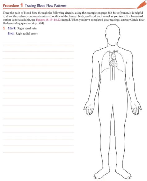
Table of Contents
- Procedure 1 Tracing Blood Flow Patterns
- Table of Contents
- Procedure 1: Tracing Blood Flow Patterns: A Comprehensive Guide
- Understanding the Fundamentals of Blood Flow
- Pressure Gradients: The Driving Force
- Vascular Resistance: The Obstacle Course
- Blood Viscosity: The Thickness Factor
- Cardiac Output: The Pumping Power
- Procedure 1: A Step-by-Step Approach to Tracing Blood Flow
- Advanced Techniques for Blood Flow Analysis
- Potential Challenges and Limitations
- Conclusion
- Latest Posts
- Latest Posts
- Related Post
Procedure 1: Tracing Blood Flow Patterns: A Comprehensive Guide
Understanding blood flow patterns is fundamental to diagnosing and treating various cardiovascular conditions. This detailed guide explores Procedure 1, a hypothetical yet comprehensive methodology encompassing various techniques for tracing blood flow. While specific procedures vary depending on the clinical setting and technology available, this article outlines the core principles and steps involved in effective blood flow pattern analysis.
Understanding the Fundamentals of Blood Flow
Before delving into the procedural steps, it's crucial to grasp the underlying principles governing blood flow. Several key factors influence the pattern and velocity of blood:
Pressure Gradients: The Driving Force
Blood flow is primarily driven by pressure gradients. Blood moves from areas of high pressure (like the left ventricle of the heart) to areas of low pressure (like the peripheral capillaries). Any obstruction or alteration in pressure significantly affects flow patterns.
Vascular Resistance: The Obstacle Course
Vascular resistance represents the opposition to blood flow within the circulatory system. Factors contributing to resistance include vessel diameter, blood viscosity, and vessel length. Narrowed arteries (stenosis), for instance, significantly increase resistance and alter flow patterns.
Blood Viscosity: The Thickness Factor
Blood viscosity refers to the thickness of blood. Higher viscosity (e.g., in conditions like polycythemia) increases resistance and slows down blood flow. Conversely, lower viscosity enhances flow.
Cardiac Output: The Pumping Power
The cardiac output, the volume of blood pumped by the heart per minute, directly influences blood flow. A stronger heart pump increases output, leading to higher flow rates throughout the system.
Procedure 1: A Step-by-Step Approach to Tracing Blood Flow
This hypothetical "Procedure 1" integrates various non-invasive and minimally invasive techniques for comprehensive blood flow analysis. Remember, specific procedures may differ based on the clinical context.
Phase 1: Initial Assessment and Patient Preparation
-
Medical History and Physical Examination: A thorough review of the patient's medical history, including symptoms, risk factors (e.g., hypertension, smoking), and family history of cardiovascular disease, is essential. A physical examination, including auscultation (listening to heart sounds) and palpation (feeling for pulses), provides initial clues about potential flow abnormalities.
-
Non-Invasive Imaging: Getting a Big Picture
-
Echocardiography: This ultrasound-based technique provides real-time images of the heart's structure and function, revealing abnormalities in valve function, chamber size, and blood flow within the heart. Doppler echocardiography specifically measures blood flow velocity and direction.
-
Doppler Ultrasound: This technique uses sound waves to assess blood flow in peripheral arteries and veins. It's valuable in detecting stenosis, thrombosis (blood clot formation), and other flow obstructions in limbs.
-
Magnetic Resonance Angiography (MRA): MRA utilizes magnetic fields and radio waves to create detailed images of blood vessels, allowing for the visualization of complex flow patterns and the detection of aneurysms or other vascular abnormalities.
-
Computed Tomography Angiography (CTA): Similar to MRA, CTA uses X-rays to produce detailed images of blood vessels. It's often faster than MRA but exposes the patient to ionizing radiation.
-
Phase 2: Targeted Investigation (If Necessary)
If the initial assessment reveals potential flow abnormalities, further investigation may be necessary:
-
Cardiac Catheterization: This minimally invasive procedure involves inserting a catheter into an artery (usually in the groin) and advancing it to the heart. It allows for direct visualization of coronary arteries, measurement of pressure gradients, and assessment of blood flow using angiography (injection of contrast dye). This is crucial for diagnosing coronary artery disease.
-
Electrocardiography (ECG): ECG records the heart's electrical activity. While not directly visualizing blood flow, ECG helps identify arrhythmias (irregular heartbeats) that can indirectly affect blood flow patterns.
-
Stress Testing: Exercise stress tests or pharmacological stress tests evaluate the heart's response to increased demand. Changes in ECG and blood pressure during stress testing can indicate impaired blood flow to the heart muscle (myocardium).
Phase 3: Data Analysis and Interpretation
-
Image Analysis: Specialized software is used to analyze the images obtained from echocardiography, Doppler ultrasound, MRA, and CTA. This involves measuring blood flow velocities, calculating pressure gradients, and assessing vessel diameter.
-
Hemodynamic Parameters: Data from cardiac catheterization, including pressure measurements and flow rates, are meticulously analyzed to assess the severity of any flow obstructions.
-
Correlation with Clinical Findings: The results of all diagnostic tests are integrated with the patient's clinical presentation and medical history to form a comprehensive diagnosis.
Phase 4: Report Generation and Treatment Planning
-
Comprehensive Report: A detailed report summarizing all findings is generated, including images, measurements, and interpretations.
-
Treatment Strategies: Based on the diagnosis, appropriate treatment strategies are developed. This could include lifestyle modifications (diet, exercise), medication (e.g., blood thinners, antihypertensives), or interventional procedures (e.g., angioplasty, stenting, bypass surgery).
Advanced Techniques for Blood Flow Analysis
Beyond the basic procedures outlined above, several advanced techniques offer enhanced precision and detail in tracing blood flow patterns:
-
4D Flow MRI: This advanced MRI technique provides a three-dimensional visualization of blood flow over time, giving a more comprehensive understanding of complex flow dynamics.
-
Phase Contrast MRI: This MRI technique measures blood velocity and flow direction with high accuracy.
Potential Challenges and Limitations
While Procedure 1 provides a robust approach to tracing blood flow patterns, several challenges and limitations exist:
-
Cost and Accessibility: Advanced imaging techniques like MRA and CTA can be expensive and may not be readily available in all healthcare settings.
-
Radiation Exposure: CTA exposes patients to ionizing radiation, which carries potential long-term health risks.
-
Patient Factors: Patient factors like obesity or claustrophobia can affect the quality of imaging studies.
-
Interpretation Complexity: Analyzing complex blood flow patterns requires expertise and experience. Accurate interpretation is crucial for appropriate diagnosis and management.
Conclusion
Tracing blood flow patterns is a crucial aspect of cardiovascular diagnostics. Procedure 1, as outlined here, represents a multi-faceted approach that integrates various techniques to provide a comprehensive understanding of blood flow dynamics. While specific procedures vary, the fundamental principles of pressure gradients, vascular resistance, blood viscosity, and cardiac output remain paramount in interpreting blood flow patterns and guiding appropriate clinical management. Continuous advancements in imaging technology and analytical techniques are improving the accuracy and efficiency of blood flow analysis, leading to better patient care and improved cardiovascular health outcomes. This detailed guide serves as a foundation for understanding the complexity and importance of precisely tracing blood flow in clinical practice. Further research and development in this field are vital to refining techniques and improving the diagnosis and treatment of cardiovascular diseases.
Latest Posts
Related Post
Thank you for visiting our website which covers about Procedure 1 Tracing Blood Flow Patterns . We hope the information provided has been useful to you. Feel free to contact us if you have any questions or need further assistance. See you next time and don't miss to bookmark.
