Preganglionic Fibers Leave The Cns And Then Synapse On
Holbox
Mar 28, 2025 · 6 min read
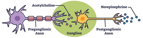
Table of Contents
- Preganglionic Fibers Leave The Cns And Then Synapse On
- Table of Contents
- Preganglionic Fibers: Leaving the CNS and Synapsing on Ganglionic Neurons
- The Autonomic Nervous System: A Brief Overview
- The Journey of Preganglionic Fibers: From CNS to Ganglia
- Sympathetic Preganglionic Fibers: The Thoracolumbar Outflow
- Parasympathetic Preganglionic Fibers: The Craniosacral Outflow
- Neurotransmitters and Receptors: A Closer Look
- Clinical Significance: Understanding Dysfunction
- Conclusion: A Complex System for Homeostasis
- Latest Posts
- Latest Posts
- Related Post
Preganglionic Fibers: Leaving the CNS and Synapsing on Ganglionic Neurons
The autonomic nervous system (ANS), a crucial component of the peripheral nervous system (PNS), regulates involuntary bodily functions like heart rate, digestion, and respiration. Understanding its intricate workings, particularly the pathways of preganglionic fibers, is essential for comprehending overall bodily homeostasis. This article delves into the journey of preganglionic fibers as they exit the central nervous system (CNS) and subsequently synapse onto ganglionic neurons, explaining the complexities of this process and its significance in maintaining bodily functions.
The Autonomic Nervous System: A Brief Overview
Before exploring preganglionic fibers, it's vital to establish a foundational understanding of the ANS. The ANS is broadly divided into two branches: the sympathetic and parasympathetic nervous systems. These branches often exert opposing effects on target organs, working in a delicate balance to maintain optimal physiological conditions.
Sympathetic Nervous System: Primarily involved in the "fight-or-flight" response, the sympathetic system prepares the body for stressful situations. It increases heart rate, blood pressure, and respiration while diverting blood flow to skeletal muscles.
Parasympathetic Nervous System: Responsible for the "rest-and-digest" functions, the parasympathetic system conserves energy and promotes restorative processes. It slows heart rate, stimulates digestion, and constricts pupils.
Both branches utilize a two-neuron pathway:
- Preganglionic neuron: Its cell body resides within the CNS, and its axon extends to synapse with a postganglionic neuron.
- Postganglionic neuron: Located in autonomic ganglia outside the CNS, its axon innervates the target organ or tissue.
The Journey of Preganglionic Fibers: From CNS to Ganglia
Preganglionic fibers, originating from the CNS, embark on a specific journey to reach their target ganglia. This journey is meticulously organized and varies slightly depending on whether the fibers belong to the sympathetic or parasympathetic nervous system.
Sympathetic Preganglionic Fibers: The Thoracolumbar Outflow
Sympathetic preganglionic neurons' cell bodies are located in the lateral horns of the spinal cord's thoracic and lumbar segments (T1-L2). This is often referred to as the thoracolumbar outflow. Their axons exit the spinal cord via the ventral roots and enter the sympathetic chain ganglia, also known as paravertebral ganglia. These ganglia form a paired chain running alongside the vertebral column.
Synaptic Options for Sympathetic Preganglionic Fibers:
Several possibilities exist for the synapse of sympathetic preganglionic fibers:
- Synapse in the same-level ganglion: The preganglionic fiber may synapse directly with a postganglionic neuron in the ganglion at the same spinal level.
- Synapse in a superior or inferior ganglion: The fiber might ascend or descend the sympathetic chain, synapsing in a ganglion at a different level. This allows for widespread coordinated responses.
- Synapse in a prevertebral ganglion: Some preganglionic fibers bypass the sympathetic chain entirely, extending to prevertebral ganglia (e.g., celiac, superior mesenteric, inferior mesenteric ganglia) located anterior to the vertebral column. These ganglia innervate abdominal viscera.
- Direct innervation of the adrenal medulla: Preganglionic fibers directly innervate the adrenal medulla, the inner portion of the adrenal gland. Instead of synapsing with postganglionic neurons, they stimulate the release of epinephrine (adrenaline) and norepinephrine (noradrenaline) into the bloodstream, producing a widespread systemic effect.
Parasympathetic Preganglionic Fibers: The Craniosacral Outflow
In contrast to the sympathetic system's thoracolumbar outflow, the parasympathetic system exhibits a craniosacral outflow. Parasympathetic preganglionic neurons originate in the brainstem (cranial nerves III, VII, IX, and X) and the sacral spinal cord (S2-S4).
Cranial Parasympathetic Outflow: The vagus nerve (CN X) carries the majority of parasympathetic preganglionic fibers, innervating numerous thoracic and abdominal organs. Other cranial nerves contribute to parasympathetic innervation of specific structures, such as the eyes and salivary glands.
Sacral Parasympathetic Outflow: Preganglionic fibers from the sacral spinal cord innervate pelvic viscera, including the bladder, rectum, and reproductive organs.
Synapse Location for Parasympathetic Preganglionic Fibers:
Parasympathetic ganglia are typically located close to or even within the walls of target organs. Therefore, parasympathetic preganglionic fibers have relatively long axons compared to their sympathetic counterparts. The synapse with the postganglionic neuron occurs in these terminal ganglia.
Neurotransmitters and Receptors: A Closer Look
The communication between preganglionic and postganglionic neurons, and subsequently between postganglionic neurons and target organs, involves specific neurotransmitters and receptors.
Acetylcholine (ACh): ACh is the primary neurotransmitter released by all preganglionic neurons, both sympathetic and parasympathetic.
Nicotinic Receptors: Postganglionic neurons possess nicotinic acetylcholine receptors (nAChRs) on their cell bodies. These receptors are ionotropic, meaning they directly open ion channels upon ACh binding, leading to depolarization and excitation of the postganglionic neuron.
Postganglionic Neurotransmitters:
- Sympathetic Postganglionic Neurons: Most sympathetic postganglionic neurons release norepinephrine (NE), which binds to adrenergic receptors (α and β receptors) on target organs. However, some sympathetic postganglionic neurons, such as those innervating sweat glands, release ACh.
- Parasympathetic Postganglionic Neurons: Parasympathetic postganglionic neurons typically release ACh, which binds to muscarinic acetylcholine receptors (mAChRs) on target organs. Muscarinic receptors are metabotropic, meaning they initiate a cascade of intracellular signaling events.
Clinical Significance: Understanding Dysfunction
Dysfunction within the pathways of preganglionic fibers can lead to various clinical manifestations. Damage to preganglionic neurons or their axons can disrupt autonomic function, resulting in conditions such as:
- Orthostatic hypotension: Inability to maintain blood pressure upon standing, often due to impaired sympathetic control of blood vessels.
- Gastrointestinal motility disorders: Problems with digestion and bowel function, potentially stemming from parasympathetic dysfunction.
- Bladder dysfunction: Difficulties with urination due to impaired parasympathetic or sympathetic innervation of the bladder.
- Horner's syndrome: A constellation of symptoms resulting from damage to sympathetic fibers innervating the head and neck, causing ptosis (drooping eyelid), miosis (pupil constriction), and anhidrosis (lack of sweating).
Conclusion: A Complex System for Homeostasis
The intricate pathways of preganglionic fibers, their synaptic connections, and the neurochemical interactions they mediate are fundamental to the proper functioning of the autonomic nervous system. Understanding this complex system is crucial not only for comprehending normal physiological processes but also for diagnosing and managing a range of clinical conditions arising from autonomic dysfunction. The detailed exploration provided in this article underscores the importance of this often-overlooked yet vital aspect of human physiology. Further research into the intricacies of preganglionic fiber pathways continues to expand our understanding of this crucial system and its role in maintaining overall health and well-being. The specific interactions between preganglionic neurons and their postganglionic counterparts, along with the downstream effects on target tissues, remain areas of active investigation, promising further advancements in our knowledge of the autonomic nervous system and related clinical conditions. The complex interplay of neurotransmitters and receptors, their specific locations and roles, all contribute to the sophisticated and finely-tuned regulation of our internal environment. A deep understanding of this system allows for better diagnostics and ultimately, more effective treatment strategies for associated disorders.
Latest Posts
Latest Posts
-
The Develpment Of Promotional Stragties Most Likely
Mar 31, 2025
-
Ige Antibodies Are Best Described As
Mar 31, 2025
-
What Is Shown In This Picture
Mar 31, 2025
-
Correctly Label The Following Anatomical Features Of The Elbow Joint
Mar 31, 2025
-
If Services Are Rendered On Account Then
Mar 31, 2025
Related Post
Thank you for visiting our website which covers about Preganglionic Fibers Leave The Cns And Then Synapse On . We hope the information provided has been useful to you. Feel free to contact us if you have any questions or need further assistance. See you next time and don't miss to bookmark.
