Pal Cadaver Axial Skeleton Skull Lab Practical Question 6
Holbox
Mar 30, 2025 · 6 min read

Table of Contents
- Pal Cadaver Axial Skeleton Skull Lab Practical Question 6
- Table of Contents
- Pal Cadaver Axial Skeleton Skull Lab Practical: Question 6 and Beyond
- Understanding the Pal Cadaver and its Importance
- The Skull: A Complex Structure
- Tackling Question 6: A Hypothetical Example
- Detailed Breakdown of Key Structures:
- Expanding Beyond Question 6: Further Exploration
- Cranial Bones: A Detailed Overview
- Facial Bones: Adding to the Complexity
- Foramina and Fissures: Passageways of Significance
- Sutures: Understanding the Joints
- Effective Study Strategies for Success
- Conclusion: Mastering the Pal Cadaver Skull
- Latest Posts
- Latest Posts
- Related Post
Pal Cadaver Axial Skeleton Skull Lab Practical: Question 6 and Beyond
This article delves deep into the complexities of Question 6 (and beyond!) in a typical pal cadaver axial skeleton skull lab practical. We'll cover crucial anatomical structures, common points of confusion, effective study strategies, and tips to ace your practical exam. Understanding the skull is fundamental to comprehending human anatomy, and this detailed guide will help you navigate its intricacies with confidence.
Understanding the Pal Cadaver and its Importance
Before we tackle Question 6 specifically, let's establish the significance of using pal cadavers in anatomical studies. Palpable cadavers, often used in medical and dental schools, provide an unparalleled opportunity for hands-on learning. Unlike diagrams or 3D models, working with a real specimen allows for a tactile understanding of bone structure, joint articulation, and anatomical variations. This practical experience is invaluable for developing diagnostic skills and solidifying theoretical knowledge. The axial skeleton, encompassing the skull, vertebral column, and rib cage, represents a critical area of focus.
The Skull: A Complex Structure
The skull, a central component of the axial skeleton, is a marvel of biological engineering. It protects the brain, houses sensory organs, and provides attachments for muscles involved in chewing, facial expression, and head movement. Its complex structure, composed of numerous bones fused together, can be daunting for students. However, by breaking it down into manageable sections and understanding the relationships between these sections, mastering the skull becomes achievable.
Tackling Question 6: A Hypothetical Example
Let's assume Question 6 in your lab practical focuses on identifying specific cranial sutures and foramina. This is a common theme, testing your knowledge of bone landmarks and their functional significance. We'll build upon this hypothetical scenario to explore key concepts and provide practical advice.
Hypothetical Question 6:
Identify and describe the location and function of the following structures on the provided pal cadaver skull: Coronal Suture, Lambdoid Suture, Foramen Magnum, and Supraorbital Foramen.
Detailed Breakdown of Key Structures:
-
Coronal Suture: This fibrous joint connects the frontal bone to the parietal bones. It's located across the top of the skull, running from ear to ear. Its function is to allow for slight movement during childbirth and to accommodate brain growth in infancy. Look for its characteristic serrated appearance on the pal cadaver.
-
Lambdoid Suture: This suture joins the occipital bone to the parietal bones. It's positioned at the back of the skull, resembling the Greek letter lambda (Λ). Similar to the coronal suture, it allows for flexibility during development. Note its location in relation to the occipital protuberance.
-
Foramen Magnum: This large opening in the occipital bone is of paramount importance, as it allows the spinal cord to pass from the brain to the vertebral column. It’s easily identifiable as the largest opening at the base of the skull. Be certain to appreciate its location and the critical structures that pass through it.
-
Supraorbital Foramen (or Notch): Located on the frontal bone, above each orbit (eye socket), this foramen (or notch – sometimes it's incomplete and forms a notch) transmits the supraorbital nerve and artery, supplying sensation and blood to the forehead and upper eyelid. Its location is crucial for understanding the distribution of the nerve supply.
Answering the Question Effectively:
When answering such a question, be systematic and precise:
-
Clear Identification: Point clearly to each structure on the pal cadaver skull. Avoid ambiguity.
-
Precise Location: Use anatomical terminology to describe the location of each structure (e.g., "located at the junction of the frontal and parietal bones").
-
Functional Significance: Explain the function of each structure, demonstrating your understanding of its role in the overall functioning of the skull.
-
Articulation: Demonstrate understanding of how the bones articulate with each other at the sutures.
-
Clinical Relevance (Optional but beneficial): If possible, briefly mention clinical relevance. For instance, premature closure of sutures (craniosynostosis) can lead to skull deformities.
Expanding Beyond Question 6: Further Exploration
While Question 6 might focus on specific structures, a comprehensive understanding of the skull requires a broader knowledge base. Here are some areas to explore further:
Cranial Bones: A Detailed Overview
-
Frontal Bone: Forms the forehead and part of the orbits.
-
Parietal Bones (2): Form the majority of the skull's superior and lateral aspects.
-
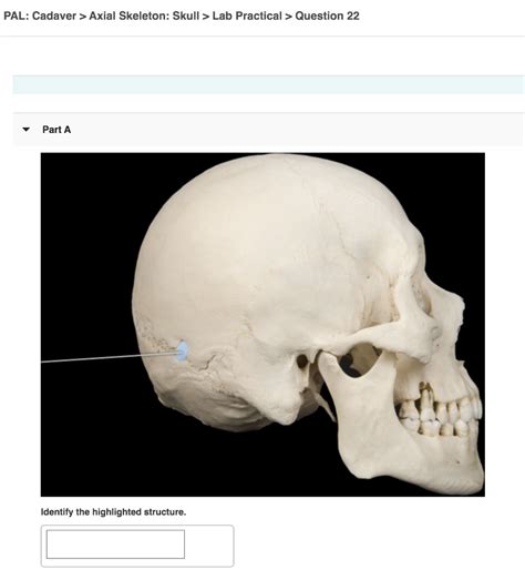
Temporal Bones (2): Located on the sides of the skull, containing the middle and inner ear structures and the mandibular fossa (jaw articulation). Identify the mastoid process and zygomatic process.
-
Occipital Bone: Forms the posterior base of the skull, including the foramen magnum.
-
Sphenoid Bone: A complex, butterfly-shaped bone forming part of the base of the skull, orbits, and nasal cavity.
-
Ethmoid Bone: Located in the anterior cranial fossa, contributing to the nasal cavity and orbits.
Facial Bones: Adding to the Complexity
The facial bones contribute significantly to the skull's overall structure and function. Mastering these bones is crucial for a thorough understanding:
-
Maxillae (2): Form the upper jaw, housing the upper teeth.
-
Mandible: The lower jaw, the only movable bone in the skull.
-
Zygomatic Bones (2): Form the cheekbones.
-
Nasal Bones (2): Form the bridge of the nose.
-
Lacrimal Bones (2): Small bones forming part of the medial wall of each orbit.
-
Vomer: Forms the posterior part of the nasal septum.
-
Inferior Nasal Conchae (2): Curved bones within the nasal cavity.
Foramina and Fissures: Passageways of Significance
Numerous foramina (openings) and fissures (clefts) allow for the passage of nerves, blood vessels, and other structures. Understanding their location and the structures they transmit is essential. Some key examples include:
-
Optic Canal: Transmits the optic nerve.
-
Superior Orbital Fissure: Transmits several cranial nerves controlling eye movements.
-
Inferior Orbital Fissure: Transmits nerves and vessels to the orbit.
-
Stylomastoid Foramen: Transmits the facial nerve.
-
Jugular Foramen: Transmits the internal jugular vein and several cranial nerves.
Sutures: Understanding the Joints
Sutures are fibrous joints connecting the bones of the skull. Understanding their location and the bones they connect is crucial. Key sutures to study beyond those mentioned in Question 6 include:
-
Squamous Suture: Connects the temporal bone to the parietal bone.
-
Sagittal Suture: Connects the two parietal bones.
-
Metopic Suture: A frontal suture which may persist (or be visible) in adults.
Effective Study Strategies for Success
To master the complexities of the skull, adopt a multifaceted learning approach:
-
Hands-on Practice: Repeatedly examine the pal cadaver skull. Trace the sutures, locate the foramina, and manipulate the bones to understand their articulations.
-
Visual Aids: Use anatomical atlases, 3D models, and online resources to supplement your hands-on experience.
-
Active Recall: Test yourself frequently. Use flashcards, practice questions, and self-quizzes to reinforce your learning.
-
Clinical Correlation: Relate the anatomical structures to their clinical significance. Understanding the consequences of fractures, infections, or other pathologies will enhance your learning.
-
Study Groups: Collaborate with fellow students to discuss challenging concepts and reinforce your understanding through peer teaching.
Conclusion: Mastering the Pal Cadaver Skull
The pal cadaver axial skeleton skull lab practical, including Question 6 and beyond, requires a dedicated and systematic approach. By understanding the individual bones, their articulations, the foramina and fissures, and their clinical relevance, you can confidently navigate the complexities of this vital anatomical region. Remember that consistent hands-on practice, coupled with effective study strategies, will be your key to success. Good luck!
Latest Posts
Latest Posts
-
Humans Carry A Variety Of Non Functional Genetic Sequences Called
Apr 02, 2025
-
Michael Is An Art Elective Programme Student
Apr 02, 2025
-
A Large Negative Gdp Gap Implies
Apr 02, 2025
-
Which Of The Techniques Are Examples Of Biotechnology
Apr 02, 2025
-
The Scores Of A Recent Test Taken By 1200
Apr 02, 2025
Related Post
Thank you for visiting our website which covers about Pal Cadaver Axial Skeleton Skull Lab Practical Question 6 . We hope the information provided has been useful to you. Feel free to contact us if you have any questions or need further assistance. See you next time and don't miss to bookmark.
