Match Each Type Of Bone Marking With Its Definition
Holbox
Mar 17, 2025 · 5 min read
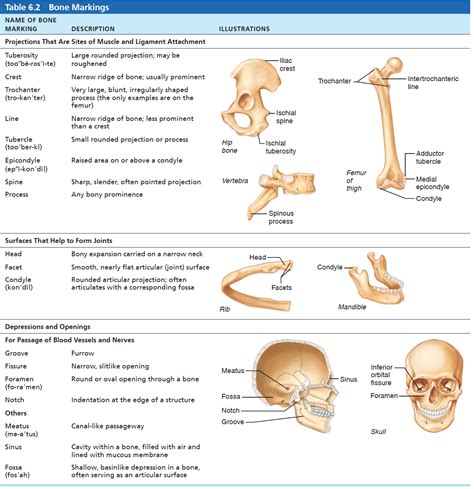
Table of Contents
Match Each Type of Bone Marking with its Definition: A Comprehensive Guide
Understanding bone markings is crucial for anyone studying anatomy, whether you're a medical student, a physical therapist, or simply an anatomy enthusiast. These markings, also known as bone features, are surface variations that reflect the stresses and strains placed on bones during growth and throughout life. They provide vital attachment points for muscles, ligaments, and tendons, and contribute significantly to the overall skeletal system's functionality. This comprehensive guide will delve into the various types of bone markings, matching each with its precise definition and providing clear examples for better understanding.
Categories of Bone Markings
Before we dive into specific markings, it’s essential to categorize them for a more structured learning experience. Bone markings can be broadly classified into two main groups:
1. Depressions and Openings: These features provide pathways for blood vessels, nerves, and ligaments, or accommodate joints.
2. Processes: These are projections or outgrowths of bone that serve as attachment points for muscles, tendons, and ligaments.
Detailed Breakdown of Bone Markings with Definitions and Examples
Now, let's explore the different types of bone markings in detail, categorized for clarity:
Depressions and Openings:
1. Fissure: A narrow, slit-like opening. Example: Superior orbital fissure (located in the sphenoid bone, allowing passage of cranial nerves and blood vessels).
2. Foramen: A rounded opening or hole. Example: Foramen magnum (located in the occipital bone, allowing the spinal cord to connect with the brain). Another example is the mental foramen in the mandible.
3. Fossa: A shallow, dish-shaped depression. Example: Mandibular fossa (located in the temporal bone, forming part of the temporomandibular joint). The olecranon fossa on the humerus is another good example.
4. Groove: A furrow or elongated depression. Example: Intertubercular groove of the humerus (accommodates the biceps brachii tendon). The radial groove is also a significant example, running along the shaft of the humerus.
5. Meatus: A tube-like passageway. Example: External acoustic meatus (located in the temporal bone, leading to the middle ear).
6. Sinus: A cavity or hollow space within a bone. Example: Paranasal sinuses (air-filled spaces within the skull bones, reducing the weight of the skull and affecting voice resonance).
7. Sulcus: A shallow groove or furrow. Example: The sulcus for the radial nerve found on the humerus. This is a common example of a sulcus running along a long bone's shaft.
Processes (Projections):
1. Condyle: A smooth, rounded articular process (a point where bone meets another bone). Example: Occipital condyles (articulate with the first cervical vertebra (atlas)). The medial and lateral condyles of the femur are also prominent.
2. Crest: A prominent, narrow, ridge-like projection. Example: Iliac crest (a prominent ridge on the ilium, providing attachment sites for muscles).
3. Epicondyle: A projection located superior to a condyle. Example: Medial and lateral epicondyles of the humerus (serving as attachment points for forearm muscles).
4. Facet: A small, flat, articular surface. Example: Articular facets of vertebrae (forming the joints between adjacent vertebrae).
5. Head: A rounded, articular projection supported on a constricted portion (neck) of a bone. Example: Head of the femur (articulates with the acetabulum of the hip bone).
6. Line: A long, narrow, slightly raised ridge. Example: Linea aspera (a rough line on the posterior surface of the femur).
7. Malleolus: A rounded process. Example: Medial malleolus of the tibia and the lateral malleolus of the fibula (form the medial and lateral projections of the ankle).
8. Protuberance: A bony prominence. Example: Mental protuberance (a bony prominence of the mandible, forming the chin).
9. Spine: A sharp, slender, pointed process. Example: Spine of the scapula (provides attachment sites for muscles).
10. Trochanter: A very large, blunt, irregularly shaped process. Example: Greater and lesser trochanters of the femur (provide attachment sites for thigh muscles).
11. Tubercle: A small, rounded process. Example: Greater tubercle of the humerus (attachment site for muscles).
12. Tuberosity: A large, rounded, roughened process. Example: Tibial tuberosity (attachment site for the patellar ligament).
Clinical Significance of Bone Markings
Understanding bone markings is not merely an academic exercise; it holds significant clinical relevance:
-
Fracture Identification and Treatment: Knowing the locations and characteristics of bone markings helps clinicians accurately diagnose and treat fractures. For instance, a fracture involving the greater trochanter of the femur requires a different treatment approach than a fracture of the shaft of the femur.
-
Surgical Procedures: Surgeons rely on their knowledge of bone markings during various procedures. For example, during hip replacement surgery, precise identification of the femoral head and neck is crucial.
-
Muscle and Ligament Attachment: Understanding the attachment points of muscles and ligaments to bone markings allows for a better understanding of the biomechanics of movement and injury mechanisms. This knowledge is especially important for physical therapists and athletic trainers.
-
Radiological Interpretation: Radiographic images (X-rays, CT scans, etc.) often reveal bone markings, helping radiologists identify underlying conditions.
Mnemonic Devices for Memorization
Memorizing the numerous bone markings can be challenging. To aid in retention, consider using mnemonic devices:
-
Categorization: Group the markings by type (depression, process, etc.).
-
Visualization: Use anatomical models or illustrations to visualize the locations and shapes of the markings.
-
Association: Create associations between the name of a marking and its function or location. For example, "condyle" sounds like "round," which reflects its shape.
-
Flashcards: Use flashcards to test yourself on the definitions and locations of the various markings.
Further Exploration
This guide provides a comprehensive overview of bone markings. For a deeper understanding, consult advanced anatomy textbooks and atlases. Interactive anatomy software and online resources can also be valuable tools for enhancing your learning.
This extensive exploration of bone markings, coupled with strategies for effective learning, equips you with the knowledge to confidently navigate the complexities of the skeletal system. By understanding the significance of these markings in both anatomical structure and clinical practice, you gain a solid foundation for further studies in anatomy, physiology, and related medical fields. Remember consistent study and practice are key to mastering this essential aspect of human anatomy.
Latest Posts
Latest Posts
-
Wordpress Is Popular Free And Open Source
Mar 17, 2025
-
The Difference Between Aerobic And Anaerobic Glucose Breakdown Is
Mar 17, 2025
-
On December 29 2020 Patel Products
Mar 17, 2025
-
A Customer Tells His Current Sales Rep
Mar 17, 2025
-
When Using A Self Managed Team A Manager Should
Mar 17, 2025
Related Post
Thank you for visiting our website which covers about Match Each Type Of Bone Marking With Its Definition . We hope the information provided has been useful to you. Feel free to contact us if you have any questions or need further assistance. See you next time and don't miss to bookmark.
