Match Each Spinal Nerve With The Main Structures It Supplies
Holbox
Mar 24, 2025 · 5 min read
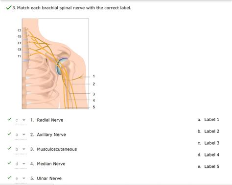
Table of Contents
- Match Each Spinal Nerve With The Main Structures It Supplies
- Table of Contents
- Matching Spinal Nerves to the Structures They Supply: A Comprehensive Guide
- Spinal Nerve Organization: A Quick Recap
- Detailed Breakdown of Spinal Nerve Innervation
- Cervical Nerves (C1-C8)
- Thoracic Nerves (T1-T12)
- Lumbar Nerves (L1-L5) & Sacral Nerves (S1-S5) (Lumbosacral Plexus)
- Coccygeal Nerve (Co1)
- Clinical Significance
- Dermatomes and Myotomes: Mapping Sensory and Motor Innervation
- Further Exploration and Resources
- Conclusion
- Latest Posts
- Latest Posts
- Related Post
Matching Spinal Nerves to the Structures They Supply: A Comprehensive Guide
Understanding the intricate network of spinal nerves and their corresponding innervation patterns is crucial for healthcare professionals, students, and anyone interested in human anatomy and physiology. This comprehensive guide meticulously details the main structures supplied by each spinal nerve, providing a detailed overview of their distribution and functions. Remember, this information is for educational purposes and should not be used for self-diagnosis or treatment. Consult a medical professional for any health concerns.
Spinal Nerve Organization: A Quick Recap
Before delving into the specific innervation patterns, let's briefly review the organization of spinal nerves. Thirty-one pairs of spinal nerves emerge from the spinal cord, named according to their vertebral level of origin:
- Cervical (C1-C8): 8 pairs, supplying the neck, shoulders, arms, and hands.
- Thoracic (T1-T12): 12 pairs, supplying the chest, abdomen, and back.
- Lumbar (L1-L5): 5 pairs, supplying the lower back, hips, and legs.
- Sacral (S1-S5): 5 pairs, supplying the buttocks, genitalia, and legs.
- Coccygeal (Co1): 1 pair, supplying a small area around the coccyx.
Each spinal nerve is formed by the union of dorsal (sensory) and ventral (motor) roots. The dorsal root carries sensory information from the periphery to the spinal cord, while the ventral root carries motor commands from the spinal cord to the muscles. These nerves then branch extensively, creating a complex network of innervation throughout the body.
Detailed Breakdown of Spinal Nerve Innervation
It's impossible to provide an exhaustive list of every structure supplied by each spinal nerve due to the extensive branching and overlapping innervation. However, we will focus on the major structures and regions supplied by each nerve group. Keep in mind that there can be considerable individual variation.
Cervical Nerves (C1-C8)
C1 (Suboccipital Nerve): Primarily supplies the muscles of the posterior neck, including the rectus capitis posterior major and minor, and obliquus capitis superior and inferior. It also contributes to proprioception in the upper neck.
C2 (Greater Occipital Nerve): Provides sensory innervation to the scalp, posterior neck, and upper part of the back of the head.
C3 & C4 (Cervical Plexus): These nerves contribute to the cervical plexus, supplying sensory innervation to the skin of the neck, shoulders, and upper chest. They also contribute to motor innervation of several neck muscles.
C5 - T1 (Brachial Plexus): These nerves form the brachial plexus, a complex network that supplies the entire upper limb. The main branches of the brachial plexus and their associated muscle innervation include:
- Axillary Nerve: Deltoid and teres minor muscles.
- Musculocutaneous Nerve: Biceps brachii, brachialis, and coracobrachialis muscles.
- Radial Nerve: Extensor muscles of the forearm and wrist, as well as the posterior compartment muscles of the arm.
- Median Nerve: Flexor muscles of the forearm (except flexor carpi ulnaris), thenar muscles of the hand, and some lumbricals.
- Ulnar Nerve: Flexor carpi ulnaris, most intrinsic hand muscles, and some lumbricals.
Thoracic Nerves (T1-T12)
The thoracic nerves are less complex than the cervical and lumbosacral plexuses. They primarily supply the muscles and skin of the thorax and abdomen:
- T1-T12 Intercostal Nerves: These nerves run along the intercostal spaces, providing sensory and motor innervation to the intercostal muscles, abdominal muscles, and skin of the thorax and abdomen. They also contribute to the innervation of the diaphragm (primarily through the phrenic nerve, which originates from C3-C5 but receives some contributions from T1-T3).
Lumbar Nerves (L1-L5) & Sacral Nerves (S1-S5) (Lumbosacral Plexus)
The lumbar and sacral nerves combine to form the lumbosacral plexus, a complex network that innervates the lower limb and pelvic region. Key branches and their innervation include:
-
Obturator Nerve: Adductor muscles of the thigh.
-
Femoral Nerve: Iliopsoas muscle, sartorius, quadriceps femoris, and pectineus muscles. It also provides sensory innervation to the anterior thigh and medial leg.
-
Sciatic Nerve (L4-S3): The largest nerve in the body. It divides into the tibial and common peroneal nerves.
- Tibial Nerve: Hamstrings, gastrocnemius, soleus, and other posterior leg and foot muscles. Provides sensory innervation to the posterior leg and sole of the foot.
- Common Peroneal Nerve: Tibialis anterior, extensor digitorum longus, peroneal muscles, and other anterior and lateral leg muscles. Provides sensory innervation to the anterior and lateral leg and dorsal foot.
-
Superior Gluteal Nerve: Gluteus medius and minimus, and tensor fasciae latae muscles.
-
Inferior Gluteal Nerve: Gluteus maximus muscle.
-
Pudendal Nerve: Muscles and skin of the perineum (external genitalia, anal sphincter, etc.).
Coccygeal Nerve (Co1)
The coccygeal nerve supplies a small area of skin over the coccyx.
Clinical Significance
Understanding the precise distribution of spinal nerves is crucial for diagnosing and treating a wide range of neurological conditions. For example:
- Radiculopathy: Compression or irritation of a spinal nerve root can cause pain, numbness, tingling, and weakness in the area supplied by that nerve. The location of symptoms helps pinpoint the affected nerve root.
- Peripheral Neuropathies: Damage to peripheral nerves can result in a variety of symptoms, depending on the affected nerve(s).
- Spinal Cord Injuries: Injuries to the spinal cord can affect the function of spinal nerves below the level of the injury, leading to paralysis or loss of sensation.
- Surgical Procedures: Knowledge of spinal nerve distribution is essential for neurosurgeons and other surgical specialists to avoid damaging nerves during procedures.
Dermatomes and Myotomes: Mapping Sensory and Motor Innervation
Dermatomes are areas of skin supplied by a single spinal nerve. Mapping dermatomes can help localize neurological lesions. Myotomes are groups of muscles innervated by a single spinal nerve. Testing myotomes helps assess the integrity of motor pathways. While overlapping innervation exists, understanding these concepts is crucial in clinical neurology.
Further Exploration and Resources
This guide provides a foundational understanding of spinal nerve innervation. For a deeper dive, explore advanced anatomy and neurology textbooks and resources. Remember to consult reliable medical sources for comprehensive and accurate information.
Conclusion
The intricate network of spinal nerves and their precise distribution to various structures throughout the body is a marvel of biological engineering. Thorough understanding of these patterns is essential for healthcare professionals, students, and anyone seeking a deeper understanding of human anatomy and physiology. This detailed exploration should serve as a valuable resource, although always consult reputable medical literature for the most current and accurate information. The body's complexity necessitates continued learning and exploration.
Latest Posts
Latest Posts
-
Identify The Three Major Modes Of Action Of Antiviral Drugs
Mar 26, 2025
-
Label The Structures Of The Urinary Tract In The Figure
Mar 26, 2025
-
Which Of The Following Is A Function Of Protein
Mar 26, 2025
-
Campaigning Its A Process Answer Key
Mar 26, 2025
-
Rn Pharmacology Online Practice 2023 B
Mar 26, 2025
Related Post
Thank you for visiting our website which covers about Match Each Spinal Nerve With The Main Structures It Supplies . We hope the information provided has been useful to you. Feel free to contact us if you have any questions or need further assistance. See you next time and don't miss to bookmark.
