Label The Structures Of The Pelvis
Holbox
Mar 19, 2025 · 6 min read
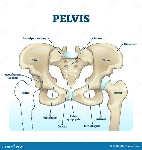
Table of Contents
Labeling the Structures of the Pelvis: A Comprehensive Guide
The pelvis, a complex bony structure located at the base of the spine, plays a crucial role in supporting the weight of the upper body, facilitating locomotion, and protecting vital organs. Understanding its intricate anatomy is essential for various fields, including medicine, physical therapy, and anatomy studies. This comprehensive guide provides a detailed description of the pelvic structures, guiding you through labeling exercises and enhancing your understanding of this fascinating region of the human body.
The Bony Pelvis: A Foundation of Support
The bony pelvis is formed by three main bones: the two hip bones (ossa coxae) and the sacrum. These bones articulate with each other to form a ring-like structure, providing stability and support.
1. Hip Bones (Ossa Coxae): A Fusion of Three
Each hip bone is actually a fusion of three separate bones during development: the ilium, ischium, and pubis.
-
Ilium: This is the largest and uppermost portion of the hip bone. Its prominent wing-like structure, the iliac crest, is easily palpable and serves as an important anatomical landmark. The anterior superior iliac spine (ASIS) and posterior superior iliac spine (PSIS) are located at the anterior and posterior ends of the iliac crest, respectively. These spines are crucial attachment points for various muscles and ligaments. The iliac fossa, a concave surface on the inner aspect of the ilium, provides attachment for the iliacus muscle. The greater sciatic notch and lesser sciatic notch are important openings on the posterior aspect of the ilium, allowing passage for nerves and blood vessels.
-
Ischium: Located inferior to the ilium, the ischium forms the lower and back part of the hip bone. The ischial tuberosity, or "sit bone," is the prominent bony prominence that bears weight when sitting. The ischial spine projects medially and superiorly, serving as an attachment point for several ligaments.
-
Pubis: The pubis forms the anterior part of the hip bone. The two pubic bones articulate at the pubic symphysis, a cartilaginous joint that allows for slight movement during childbirth. The superior and inferior pubic rami extend laterally from the pubic body. The pubic tubercle is a small, roughened projection located on the superior ramus.
2. Sacrum: The Keystone of the Pelvic Ring
The sacrum is a triangular-shaped bone formed by the fusion of five sacral vertebrae. It articulates with the ilium at the sacroiliac joints (SIJs), strong, relatively immobile joints that are crucial for transmitting weight from the spine to the pelvis. The sacral promontory is the anterior projection of the superior border of the sacrum, serving as an important landmark in obstetrics. The sacral foramina are openings on the lateral aspects of the sacrum that transmit spinal nerves. The sacral canal is the continuation of the vertebral canal, housing the cauda equina. The apex of the sacrum articulates with the coccyx.
3. Coccyx: The Tailbone
The coccyx is a small, triangular bone formed by the fusion of three to five coccygeal vertebrae. It represents the vestigial remnant of a tail.
Important Pelvic Ligaments: Stability and Support
Numerous ligaments contribute to the stability of the pelvic girdle, reinforcing the joints and limiting excessive movement. Some of the most important ligaments include:
-
Sacroiliac ligaments: These ligaments connect the sacrum to the ilium, reinforcing the sacroiliac joints. They include the anterior sacroiliac ligament, posterior sacroiliac ligament, and interosseous sacroiliac ligament.
-
Iliolumbar ligament: This ligament connects the transverse processes of the fifth lumbar vertebra to the iliac crest.
-
Sacrotuberous ligament: This ligament extends from the sacrum and coccyx to the ischial tuberosity.
-
Sacrospinous ligament: This ligament connects the sacrum and coccyx to the ischial spine.
-
Pubic symphysis ligaments: These ligaments reinforce the pubic symphysis.
Pelvic Openings and Foramina: Pathways for Vessels and Nerves
The pelvis contains several openings and foramina that allow for the passage of blood vessels, nerves, and other structures.
-
Obturator foramen: This large opening is formed by the ischium and pubis. It is partially closed by the obturator membrane, through which the obturator nerve and vessels pass.
-
Greater sciatic foramen: This large opening is located between the greater sciatic notch and the sacrospinous and sacrotuberous ligaments. It allows for the passage of the sciatic nerve and other important structures.
-
Lesser sciatic foramen: Located inferior to the greater sciatic foramen, it transmits the internal pudendal vessels and pudendal nerve.
Pelvic Floor Muscles: Essential for Support and Function
The pelvic floor muscles form a sling-like structure that supports the pelvic organs and plays a crucial role in continence and sexual function. These muscles include the levator ani (pubococcygeus, puborectalis, iliococcygeus) and coccygeus muscles. Damage to these muscles can lead to pelvic organ prolapse or urinary incontinence.
Pelvic Inlet and Outlet: Defining the Pelvic Boundaries
The pelvis is divided into the pelvic inlet and pelvic outlet.
-
Pelvic Inlet: This is the superior opening of the bony pelvis, delineated by the sacral promontory, arcuate line of the ilium, pectineal line of the pubis, and pubic symphysis. Its shape and size are important in obstetrics.
-
Pelvic Outlet: This is the inferior opening of the bony pelvis, bounded by the pubic arch, ischial tuberosities, and coccyx. Its size is also relevant to childbirth.
Clinical Significance: Understanding Pelvic Pathology
Understanding the anatomy of the pelvis is crucial in diagnosing and treating various conditions, including:
-
Pelvic fractures: Fractures of the pelvic bones can result from high-impact trauma.
-
Sacroiliac joint dysfunction: Pain and dysfunction in the sacroiliac joints can cause low back pain.
-
Pelvic organ prolapse: This condition involves the descent of pelvic organs, such as the uterus or bladder, into the vagina.
-
Urinary incontinence: This is the involuntary leakage of urine.
-
Pelvic inflammatory disease (PID): An infection of the female reproductive organs.
Practical Labeling Exercises: Strengthening Your Knowledge
To solidify your understanding of pelvic structures, engage in active labeling exercises. You can use anatomical models, diagrams, or atlases as references. Focus on accurately identifying the bones, ligaments, foramina, and muscles. Start with labeling the major bones (ilium, ischium, pubis, sacrum, coccyx) and then progressively add more intricate structures. Use flashcards or quizzes to test your knowledge and identify areas requiring further study. Consider creating your own detailed diagrams to enhance your learning experience.
Conclusion: Mastering Pelvic Anatomy
The pelvis is a region of remarkable complexity and functional significance. Mastering the anatomy of this region requires dedicated effort and consistent practice. By systematically studying the bones, ligaments, muscles, and other structures, you'll build a strong foundation for understanding the biomechanics of the pelvis and its role in overall human health. The provided labeling exercises and detailed descriptions serve as a powerful tool in your journey towards mastering this intricate and essential part of the human body. Remember to supplement this guide with further reading and visual aids for a comprehensive understanding. Consistent review and practical application will solidify your knowledge and enhance your appreciation for the marvelous architecture of the human pelvis.
Latest Posts
Latest Posts
-
What Should You Assess Regardless Of Age Group
Mar 20, 2025
-
Assignment 7 Project Stem Leapp Year
Mar 20, 2025
-
Work For The Same Employer And Often Have Lunch Together
Mar 20, 2025
-
Which Choice Is The Best Definition Of Genetic Determinism
Mar 20, 2025
-
The Functional Filtration Unit Of The Kidney Is The
Mar 20, 2025
Related Post
Thank you for visiting our website which covers about Label The Structures Of The Pelvis . We hope the information provided has been useful to you. Feel free to contact us if you have any questions or need further assistance. See you next time and don't miss to bookmark.
