Label The Photomicrograph Of Thick Skin
Holbox
Mar 23, 2025 · 6 min read
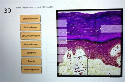
Table of Contents
- Label The Photomicrograph Of Thick Skin
- Table of Contents
- Labeling a Photomicrograph of Thick Skin: A Comprehensive Guide
- Understanding Thick Skin Structure
- 1. A Significantly Thicker Stratum Corneum:
- 2. Prominent Stratum Lucidum:
- 3. Well-Defined Strata:
- 4. Dermal Papillae and Ridges:
- 5. Thick Dermis:
- Key Structures to Label in Your Photomicrograph:
- Epidermis:
- Dermis:
- Other Structures (Potentially Visible):
- Practical Tips for Labeling:
- Beyond Labeling: Clinical Significance
- Conclusion:
- Latest Posts
- Latest Posts
- Related Post
Labeling a Photomicrograph of Thick Skin: A Comprehensive Guide
Identifying the diverse structures within a photomicrograph of thick skin requires a meticulous approach and a solid understanding of histological features. This comprehensive guide will walk you through the process, explaining the key components and their characteristic appearances. Mastering this skill is crucial for anyone studying histology, dermatology, or related fields.
Understanding Thick Skin Structure
Before we delve into labeling, it's vital to understand the unique characteristics of thick skin. Unlike thin skin, which covers most of the body, thick skin is found only on the palms of the hands and soles of the feet. This difference reflects its specialized function: providing robust protection against friction and mechanical stress. This increased protection is achieved through several key structural features:
1. A Significantly Thicker Stratum Corneum:
The stratum corneum, the outermost layer of the epidermis, is dramatically thicker in thick skin than in thin skin. This layer consists of numerous layers of dead, keratinized cells (corneocytes) that are tightly packed together, forming a tough, waterproof barrier. Its thickness is directly related to the increased protection against abrasion and environmental factors. Look for a broad, pale-staining zone in your photomicrograph, representing this dense layer of corneocytes.
2. Prominent Stratum Lucidum:
A distinct feature of thick skin is the presence of a clearly visible stratum lucidum. This translucent layer lies between the stratum corneum and stratum granulosum. It’s composed of flattened, densely packed keratinocytes with eleidin, a precursor to keratin. Under the microscope, it appears as a homogenous, eosinophilic band. Its presence is a key differentiating factor between thick and thin skin.
3. Well-Defined Strata:
While all epidermal layers are present, the other strata (stratum granulosum, stratum spinosum, and stratum basale) are also generally more organized and clearly defined in thick skin compared to thin skin.
4. Dermal Papillae and Ridges:
The dermis, the underlying connective tissue layer, shows a characteristic pattern of dermal papillae. These upward projections of the dermis interdigitate with the epidermis, increasing the surface area of contact and strengthening the dermal-epidermal junction. In thick skin, this interdigitation leads to the formation of prominent epidermal ridges (friction ridges), which are visible to the naked eye as fingerprints and footprints. Look for these characteristic downward projections of the epidermis into the dermal papillae.
5. Thick Dermis:
The dermis itself is typically thicker in thick skin than in thin skin, providing additional support and cushioning. This thicker dermis comprises both the papillary dermis (superficial, loose connective tissue) and the reticular dermis (deeper, dense irregular connective tissue). The reticular dermis is composed of densely packed collagen and elastic fibers, contributing to the skin's strength and elasticity.
Key Structures to Label in Your Photomicrograph:
Now, let's focus on the specific structures you should be able to identify and label in a typical photomicrograph of thick skin:
Epidermis:
- Stratum Corneum: The thickest layer, appearing as a pale-staining, eosinophilic zone. Note the tightly packed, anucleated keratinocytes.
- Stratum Lucidum: A thin, translucent layer located just below the stratum corneum. It appears homogenous and eosinophilic.
- Stratum Granulosum: A layer of flattened cells containing keratohyalin granules (appearing as basophilic granules). These granules are involved in keratinization.
- Stratum Spinosum: A layer of polygonal cells connected by desmosomes, giving them a spiny appearance. The cells are larger than those in the granulosum layer.
- Stratum Basale: The deepest layer of the epidermis, a single layer of cuboidal or columnar cells resting on the basement membrane. This layer contains actively dividing keratinocytes (mitotic figures may be visible) and melanocytes.
Dermis:
- Papillary Dermis: The superficial layer of the dermis, characterized by loose connective tissue, thin collagen fibers, and numerous blood vessels. This layer lies directly beneath the epidermis.
- Reticular Dermis: The deeper layer of the dermis, composed of dense irregular connective tissue with thicker collagen fibers and elastic fibers. It provides strength and elasticity to the skin.
- Dermal Papillae: Upward projections of the dermis into the epidermis. They are responsible for the formation of epidermal ridges.
- Epidermal Ridges: Downward projections of the epidermis that interdigitate with the dermal papillae. These are responsible for fingerprints and footprints.
- Hair Follicles (absent): Unlike thin skin, thick skin typically lacks hair follicles, sebaceous glands, and arrector pili muscles. The absence of these structures is a key distinguishing feature.
Other Structures (Potentially Visible):
- Meissner's Corpuscles: These are touch receptors found in the dermal papillae. They are oval-shaped and are often situated just beneath the epidermis.
- Basement Membrane: The thin layer separating the epidermis and dermis. It provides structural support and acts as a selective barrier.
- Blood Vessels: These are visible within both the papillary and reticular dermis and supply nutrients to the skin. They may appear as circular or ovoid structures.
- Nerves: Nerve fibers are present throughout the dermis, although they might not be readily apparent in all photomicrographs.
Practical Tips for Labeling:
- Use a high-quality image: A clear, well-stained photomicrograph is essential for accurate identification.
- Start with the obvious: Begin by identifying the easily recognizable layers (stratum corneum, stratum lucidum, dermis).
- Use a systematic approach: Work your way from the outermost layer (stratum corneum) to the innermost (stratum basale) and then to the dermis.
- Refer to histological diagrams: Use textbooks or online resources to compare your photomicrograph to known diagrams.
- Pay attention to staining patterns: Different layers and structures stain differently (e.g., eosinophilic, basophilic).
- Practice makes perfect: The more photomicrographs you label, the better you'll become at identifying structures.
Beyond Labeling: Clinical Significance
Understanding the histology of thick skin extends beyond academic pursuits. It has significant clinical implications:
- Wound Healing: The thicker epidermis and dermis influence wound healing processes.
- Skin Diseases: Many skin diseases present differently in thick skin compared to thin skin.
- Dermatopathology: Accurate interpretation of thick skin biopsies is vital for diagnosing skin cancers and other conditions.
- Forensic Science: Fingerprints, based on the unique ridge patterns of thick skin, are essential in forensic identification.
Conclusion:
Labeling a photomicrograph of thick skin requires careful observation and a thorough understanding of its histological features. This comprehensive guide provides a roadmap for accurately identifying and labeling the key structural components, thereby enhancing your knowledge and skills in histology and related fields. Remember that practice is key; the more you examine and label these images, the more proficient you'll become in recognizing the intricate details of this essential skin type. This improved understanding opens doors to further exploration of dermatological processes and applications. Remember to consult reliable histological textbooks and online resources for additional reference materials and further refine your ability to analyze photomicrographs.
Latest Posts
Latest Posts
-
Label The Parts Of The Hair And Hair Follicle
Mar 26, 2025
-
A Public Opinion Poll In Ohio Wants To Determine
Mar 26, 2025
-
Which Item Does Not Have Food Contact Surface
Mar 26, 2025
-
The Empirical Method Of Study Is Based On
Mar 26, 2025
-
Do Not Include The Spectating Cation
Mar 26, 2025
Related Post
Thank you for visiting our website which covers about Label The Photomicrograph Of Thick Skin . We hope the information provided has been useful to you. Feel free to contact us if you have any questions or need further assistance. See you next time and don't miss to bookmark.
