Label The Photomicrograph Of Compact Bone
Holbox
Mar 14, 2025 · 6 min read
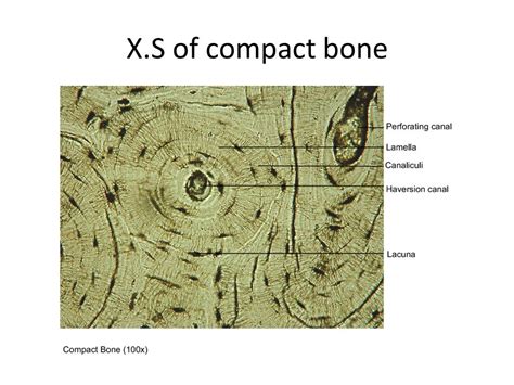
Table of Contents
Labeling the Photomicrograph of Compact Bone: A Comprehensive Guide
Understanding the microscopic structure of compact bone is crucial for anyone studying biology, anatomy, or related fields. This detailed guide will walk you through the process of accurately labeling a photomicrograph of compact bone, explaining the key structures and their functions. We'll cover everything from the fundamental building blocks to the intricate organization that gives compact bone its remarkable strength and resilience.
The Fundamental Units: Osteons (Haversian Systems)
The most striking feature of compact bone when viewed under a microscope is the presence of osteons, also known as Haversian systems. These cylindrical structures are the basic functional units of compact bone. Each osteon is composed of several key components:
1. Central Canal (Haversian Canal):
- Function: This central channel runs longitudinally through the osteon and contains blood vessels and nerves that supply the bone tissue with nutrients and oxygen, and remove waste products. Its presence is essential for maintaining the viability of the bone cells.
- Identification: On a photomicrograph, it appears as a relatively large, circular or oval space in the center of each osteon. It's often darker or lighter than the surrounding bone matrix, depending on the staining technique used.
2. Lamellae:
- Function: Concentric lamellae are rings of bone matrix surrounding the central canal. These layers are composed of collagen fibers arranged in a highly organized, spiral pattern. This specific arrangement provides exceptional strength and resistance to stress. Interstitial lamellae are remnants of older osteons that have been partially resorbed during bone remodeling. Circumferential lamellae are located around the outer and inner surfaces of the compact bone.
- Identification: Concentric lamellae are easily visible as concentric rings around the central canal. Interstitial lamellae appear as fragments of lamellae between osteons. Circumferential lamellae form a continuous layer around the entire bone. They often appear as larger, less regularly shaped bands compared to the concentric lamellae.
3. Lacunae:
- Function: These are small, spaces within the bone matrix where osteocytes, mature bone cells, reside. Each lacuna houses a single osteocyte.
- Identification: Lacunae appear as small, dark cavities within the lamellae. They are often arranged in a regular pattern, following the concentric rings of the lamellae.
4. Canaliculi:
- Function: These are extremely fine, microscopic canals that radiate from the lacunae to connect neighboring lacunae, and ultimately to the central canal. They provide pathways for the exchange of nutrients, waste products, and signaling molecules between osteocytes and the blood supply.
- Identification: Canaliculi are often difficult to see on a low-power photomicrograph but may appear as very fine lines radiating from the lacunae towards the central canal or other lacunae. Higher magnification is typically needed for clear visualization.
5. Osteocytes:
- Function: These are mature bone cells responsible for maintaining the bone matrix. They are crucial for bone remodeling and sensing mechanical stress.
- Identification: Osteocytes themselves are usually too small to be clearly visible on a typical photomicrograph. Their presence is inferred from the lacunae they occupy.
Beyond the Osteon: Other Important Structures
While osteons dominate the image of compact bone, several other structures contribute to its overall architecture and function:
1. Volkmann's Canals (Perforating Canals):
- Function: These canals run perpendicular to the central canals, connecting different osteons and supplying blood vessels and nerves to the deeper layers of compact bone. They establish a three-dimensional network of vascularization within the bone.
- Identification: Volkmann's canals appear as larger, transverse canals that intersect the osteons. They typically don't have the concentric lamellae characteristic of osteons.
2. Cement Lines:
- Function: These are thin, dark lines that mark the boundaries between adjacent osteons or between older and newer bone tissue. They represent areas of previous bone remodeling activity.
- Identification: They appear as distinct, dark lines separating the concentric lamellae of osteons or separating different osteons from each other.
3. Outer and Inner Circumferential Lamellae:
- Function: These lamellae are located at the outer and inner surfaces of the compact bone, respectively. They provide structural support and contribute to the overall strength of the bone.
- Identification: These lamellae appear as broad concentric layers located at the periphery of the compact bone section.
Practical Tips for Labeling a Photomicrograph
When labeling a photomicrograph of compact bone, use clear, concise labels. Consider the following:
- Magnification: Always include the magnification of the image. This is crucial for providing context to the size and scale of the structures observed.
- Consistent Style: Maintain a consistent style for your labels. Use the same font size and style for all labels.
- Placement: Place labels in a manner that does not obscure the underlying structures. Use arrows to point directly to the specific features you're identifying.
- Clarity: Keep your labels short, descriptive, and unambiguous. Avoid using technical jargon unless absolutely necessary.
- Accuracy: Ensure the accuracy of your labels. Double-check your identifications against reliable anatomical resources.
Applying Knowledge: Interpreting Variations
Remember that bone tissue is dynamic, constantly undergoing remodeling. Therefore, the appearance of compact bone in a photomicrograph can vary based on age, health, and location in the skeleton. You may encounter variations in:
- Osteon density: The number of osteons per unit area can vary depending on the bone's location and the stresses it experiences.
- Lamellae thickness: The width of the lamellae can vary depending on bone age and remodeling activity.
- Presence of resorption cavities: Areas of bone resorption (breakdown) may be visible, indicating bone remodeling.
- Presence of osteoclasts: These large, multinucleated cells responsible for bone resorption might be visible, particularly in areas of remodeling.
Beyond the Microscopic: Linking Structure to Function
The intricate structure of compact bone is intimately linked to its function. The organized arrangement of lamellae, the presence of osteons and canaliculi, all contribute to compact bone’s exceptional properties:
- Strength: The layered structure and spiral arrangement of collagen fibers give compact bone exceptional strength and resistance to compressive, tensile, and torsional forces.
- Lightweight: The porous nature of the bone, while providing strength, also keeps it remarkably lightweight. This is crucial for locomotion and overall skeletal efficiency.
- Dynamic Remodeling: The continuous process of bone remodeling allows for adaptation to changing mechanical loads and repair of micro-fractures.
By understanding the microscopic structure of compact bone and mastering the skill of labeling its components, you develop a deeper appreciation for the complexity and ingenuity of the human body. This knowledge is essential for anyone working in fields related to biology, anatomy, medicine, and related disciplines. The careful examination and labeling of photomicrographs offer a crucial link between theoretical understanding and practical application in the biological sciences. Remember to always utilize reputable sources and anatomical texts to confirm your identifications. Precise and accurate labeling is key to successfully communicating the intricacies of compact bone.
Latest Posts
Latest Posts
-
A Constraint In A Decision Is Blank
Mar 15, 2025
-
The Goal Of Is To Share Resources Especilaly
Mar 15, 2025
-
How Are Diploid Cells Homologous Chromosomes And Alleles Related
Mar 15, 2025
-
Early Americans Preference For Limited Government Was Strengthened By
Mar 15, 2025
-
Are Those Who Are Resonsible For Managing Large
Mar 15, 2025
Related Post
Thank you for visiting our website which covers about Label The Photomicrograph Of Compact Bone . We hope the information provided has been useful to you. Feel free to contact us if you have any questions or need further assistance. See you next time and don't miss to bookmark.
