Label The Photomicrograph Based On The Hints Provided
Holbox
Mar 15, 2025 · 5 min read
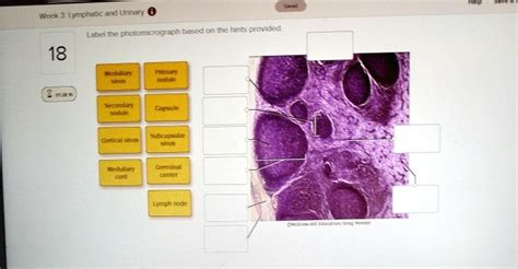
Table of Contents
Mastering Photomicrograph Labeling: A Comprehensive Guide
Photomicrography, the art of capturing images through a microscope, opens a window into the microscopic world. However, a photomicrograph is only truly valuable when correctly labeled and interpreted. This comprehensive guide will equip you with the knowledge and skills to accurately label photomicrographs, utilizing various hints and contextual information. We'll cover everything from basic labeling techniques to advanced strategies for complex images.
Understanding the Importance of Accurate Labeling
Before delving into the specifics, let's emphasize the critical role accurate labeling plays. A well-labeled photomicrograph serves several vital purposes:
- Clear Communication: It ensures your observations and findings are easily understood by others, facilitating effective communication within the scientific community or among colleagues.
- Data Integrity: Accurate labeling maintains the integrity of your scientific data, preventing misinterpretations and ensuring reproducibility of your results.
- Record Keeping: It provides a clear and permanent record of your observations, crucial for future reference and analysis.
- Educational Purposes: In educational settings, correctly labeled photomicrographs serve as valuable teaching tools, enhancing learning and understanding.
Essential Elements of a Photomicrograph Label
A complete photomicrograph label typically includes the following key elements:
- Specimen Identification: Precisely identify the organism or material being viewed. This includes the species name (genus and species), if known, and any relevant strain or variety information. For example, Escherichia coli strain DH5α.
- Magnification: Clearly state the magnification used, including both the objective lens and eyepiece magnification (e.g., 100x oil immersion).
- Staining Method (if applicable): Specify any staining techniques employed to highlight specific structures (e.g., Gram stain, Hematoxylin and Eosin stain). Include details like staining time and concentration if relevant.
- Date of Observation: Document the date the photomicrograph was taken.
- Microscopical Technique: Indicate the type of microscopy used (e.g., bright-field, phase-contrast, fluorescence, electron microscopy).
- Other Relevant Information: This might include specific experimental conditions, the preparation technique used (e.g., smear preparation, tissue sectioning), or any other pertinent observations.
Strategies for Labeling Based on Provided Hints
Now, let's delve into practical strategies for labeling photomicrographs, utilizing various hints as guides. The effectiveness of your labeling will depend significantly on the type of hints provided.
1. Hints Based on Visual Cues:
Let's assume you're presented with a photomicrograph and some visual clues. For example:
- Hint 1: "Observe the presence of cell walls and chloroplasts."
- Hint 2: "Note the characteristic shape and arrangement of the cells."
- Hint 3: "Identify any specialized structures or organelles."
Approach:
- Careful Observation: Begin by carefully scrutinizing the photomicrograph. Look for structures mentioned in the hints.
- Structure Identification: Identify the structures mentioned in the hints. For example, if the hint mentions "cell walls and chloroplasts," locate these structures within the cells.
- Shape and Arrangement: Analyze the shape and arrangement of cells. Are they spherical, rod-shaped, or filamentous? Are they arranged in chains, clusters, or individually?
- Specialized Structures: Look for any specialized structures, such as flagella, cilia, or vacuoles.
- Labeling: Label the identified structures clearly and accurately on the photomicrograph, using labels and arrows. Include a legend to clarify the meaning of each label.
Example Label:
- Specimen Identification: Elodea canadensis (Canadian waterweed)
- Magnification: 400x
- Staining Method: None
- Date of Observation: October 26, 2023
- Microscopical Technique: Bright-field microscopy
- Label: Cell wall, Chloroplast, Nucleus, Cytoplasm
2. Hints Based on Contextual Information:
Sometimes, the hints provided are not just visual cues but also contextual information. For instance:
- Hint 1: "The sample was taken from a pond water sample."
- Hint 2: "The organism is known to be photosynthetic."
- Hint 3: "Expect to see unicellular organisms."
Approach:
- Understanding the Context: Analyze the provided contextual information. The information about the pond water sample suggests that the organism might be an alga or protozoan. The information on photosynthesis narrows down the possibilities.
- Microscopic Examination: Observe the photomicrograph for features consistent with the contextual information. Look for chloroplasts (indicating photosynthesis), cell walls, and the expected morphology of unicellular organisms.
- Identifying the Organism: Try to identify the specific organism based on its morphology and the contextual information. Refer to biological resources or databases if needed.
- Labeling: Label the identified organism and its key structures.
Example Label:
- Specimen Identification: Euglena sp. (Euglena species)
- Magnification: 400x
- Staining Method: None
- Date of Observation: November 15, 2023
- Microscopical Technique: Bright-field microscopy
- Labels: Cell membrane, Chloroplast, Nucleus, Flagellum, Eyespot
3. Hints Based on Experimental Procedures:
The hints might also describe the experimental procedures employed. For instance:
- Hint 1: "Gram stain was performed on a bacterial sample."
- Hint 2: "The sample was cultured on nutrient agar."
- Hint 3: "The bacteria were known to be rod-shaped."
Approach:
- Procedure Analysis: Understand the experimental procedures described. A Gram stain differentiates between Gram-positive and Gram-negative bacteria based on cell wall characteristics. Nutrient agar is a common growth medium for bacteria.
- Interpreting the Results: The Gram stain result (Gram-positive or Gram-negative) will greatly influence your labeling. The description of rod shape will guide your observation.
- Identifying the Bacteria: Observe the photomicrograph for the morphological characteristics consistent with the Gram stain result and the description provided.
- Labeling: Label the bacteria, indicating whether they are Gram-positive or Gram-negative. Label relevant structures like cell walls, cell membranes, and any visible internal structures.
Example Label:
- Specimen Identification: Bacillus subtilis (Gram-positive bacterium)
- Magnification: 1000x oil immersion
- Staining Method: Gram stain
- Date of Observation: December 8, 2023
- Microscopical Technique: Bright-field microscopy
- Labels: Cell wall, Cell membrane, Cytoplasm, Spore (if present)
4. Advanced Labeling Techniques for Complex Photomicrographs:
For complex photomicrographs showing multiple structures or organisms, a more structured approach is necessary.
- Multiple Labels: Use multiple labels and arrows to identify each structure clearly.
- Color-Coding: Use different colors for labels to distinguish between different structures or organisms.
- Scale Bar: Include a scale bar to indicate the size of the structures in the photomicrograph.
- Detailed Legend: Provide a detailed legend explaining each label and its significance.
- Image Annotation Software: Use image annotation software for more precise labeling and measurement tools.
Conclusion:
Labeling photomicrographs accurately is a crucial skill for anyone working with microscopy. By understanding the essential elements of a label and adopting appropriate strategies based on the provided hints, you can ensure your photomicrographs are informative, accurate, and effectively communicate your scientific findings. Remember that practice is key, so keep practicing and refining your labeling techniques to master this important skill. Always strive for clarity, precision, and comprehensive labeling to maximize the value and impact of your microscopic images.
Latest Posts
Latest Posts
-
How Many Hours Are In 3 Weeks
May 19, 2025
-
How Many Feet Is 48 In
May 19, 2025
-
How Many Ml Is 8 Cups
May 19, 2025
-
95 Kilos Is How Many Pounds
May 19, 2025
-
How Many Pounds In 82 Kg
May 19, 2025
Related Post
Thank you for visiting our website which covers about Label The Photomicrograph Based On The Hints Provided . We hope the information provided has been useful to you. Feel free to contact us if you have any questions or need further assistance. See you next time and don't miss to bookmark.